<< Chapter < Page Chapter >> Page >

Sample preparation and starting the instrument

Most of proteins and peptides will require using buffers in order to prevent denaturation. Caution should be shown to avoid using any optically active buffers. Clear solutions are required. CD is taken in high transparency quartz cuvettes to ensure least interference. There are cuvettes available that have path-length ranging from 0.01 cm to 1 cm. Depending on UV activity of buffers used one should choose a cuvette with path-length (distance the beam of light passes through the sample) that compensates for UV absorbance of buffer. Solutions should be prepared according to cuvette that will be used, see [link] .

Choosing the appropriate cuvette based upon the sample concentration.
Cuvette path (cm) Concentration of sample (mg/mL)
0.01 - 0.02 0.2 – 1.0
0.1 0.05 – 0.2
1 0.005 – 0.01

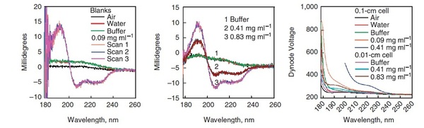
Besides, just like salts used to prepare pallets in FT-IR, the buffers in CD will show cutoffs at a certain point in low wavelength region, meaning that buffers start to absorb after certain wavelengh. The cutoff values for most of common buffers are known and can be found from manufacturer. Oxygen absorbs light below 200 nm. Therefore, in order to remove interference buffers should be prepared from distilled water or the water should be degassed before use. Another important point is to accurately determine concentration of sample, because concentration should be known for CD data analysis. Concentration of sample can be determined from extinction coefficients, if such are reported in literature also for protein samples quantitative amino acid analysis can be used.

Many CD instrument come bundled with a sample compartment temperature control unit. This is very handy when doing stability and unfolding/denaturation studies of proteins. Check to make sure the heat sink is filled with water. Turn the temperature control unit on and set to chosen temperature.

UV source in CD is very powerful lamp and can generates large amounts of Ozone in its chamber. Ozone significantly reduces the life of the lamp. Therefore, oxygen should be removed before turning on the main lamp (otherwise it will be converted to ozone near lamp). For this purpose nitrogen gas is constantly flushed into lamp compartment. Let Nitrogen flush at least for 15 min. before turning on the lamp.

Collecting spectra for blank, water, buffer background, and sample

  1. Collect spectrum of air blank ( [link] ). This will be essentially a line lying on x –axis of spectrum, zero absorbance.
  2. Fill the cuvette with water and take a spectrum.
  3. Water droplets left in cuvette may change concentration of your sample, especially when working with dilute samples. Hence, it is important to thoroughly dry the cuvette. After drying the cuvette, collect spectrum of buffer of exactly same concentration as used for sample ( [link] ). This is the step where buffer is confirmed to be suitable spectrum of the buffer and water should overlap within experimental error, except for low wavelength region where signal-to-noise ratio is low.
  4. Clean the cuvette as described above and fill with sample solution. Collect the CD spectrum for three times for better accuracy ( [link] ). For proteins multiple scans should overlap and not drift with time.
CD spectra of blank and water (left), buffer (center), and sample (right). Lysozyme in 10 mM sodium phosphate pH 7. Adapted by permission from N. Greenfield, Nat. Protoc. , 2006, 1 , 6.

Data handling and analysis

After saving the data for both the spectra of the sample and blank is smoothed using built-in commands of controller software. The smoothed baseline is subtracted from the smoothed spectrum of the sample. The next step is to use software bundles which have algorithms for estimating secondary structure of proteins. Input the data into the software package of choice and process it. The output from algorithms will be the percentage of a particular secondary structure conformation in sample. The data shown in [link] lists commonly used methods and comparers them for several proteins. The estimated secondary structure is compared to X-ray data, and one can see that it is best to use several methods for best accuracy.

Comparison of secondary structure estimation methods. Adapted by permission from N. Greenfield, Nat. Protoc. , 2006, 1 , 6.

Conclusion

What advantages CD has over other analysis methods? CD spectroscopy is an excellent, rapid method for assessing the secondary structure of proteins and performing studies of dynamic systems like folding and binding of proteins. It worth noting that CD does not provide information about the position of those subunits with specific conformation. However, CD outrivals other techniques in rapid assessing of the structure of unknown protein samples and in monitoring structural changes of known proteins caused by ligation and complex formation, temperature change, mutations, denaturants. CD is also widely used to juxtapose fused proteins with wild type counterparts, because CD spectra can tell whether the fused protein retained the structure of wild type or underwent changes.

Bibliography

  • L. Que, Physical Methods in Bioinorganic Chemistry – Spectroscopy and Magnetism , University Science Books, Sausalito (2000).
  • J. P, Wang, J. Nian, L. Xiaoqi, X. Yunhao, and C. S. Hanke, New J. Phys. , 2010, 12 , 063032.
  • N. Greenfield, Nat. Protoc. , 2006, 1 , 6.
  • G. Holzwarth and P. Doty, J. Am. Chem. Soc. , 1965, 87 , 218.
  • N. Greenfield and G.D. Fasman, Biochemistry , 1969, 8 , 4108.
  • P. Atkins and J. de Paula, Elements of Physical Chemistry , 4th ed, Oxford University Press (2005).
  • M. Rutherfurd and M. Dunn, Curr. Protoc. Protein Sci. , 2011, 63 , 3.2.1.

Questions & Answers

how does Neisseria cause meningitis
Nyibol Reply
what is microbiologist
Muhammad Reply
what is errata
Muhammad
is the branch of biology that deals with the study of microorganisms.
Ntefuni Reply
What is microbiology
Mercy Reply
studies of microbes
Louisiaste
when we takee the specimen which lumbar,spin,
Ziyad Reply
How bacteria create energy to survive?
Muhamad Reply
Bacteria doesn't produce energy they are dependent upon their substrate in case of lack of nutrients they are able to make spores which helps them to sustain in harsh environments
_Adnan
But not all bacteria make spores, l mean Eukaryotic cells have Mitochondria which acts as powerhouse for them, since bacteria don't have it, what is the substitution for it?
Muhamad
they make spores
Louisiaste
what is sporadic nd endemic, epidemic
Aminu Reply
the significance of food webs for disease transmission
Abreham
food webs brings about an infection as an individual depends on number of diseased foods or carriers dully.
Mark
explain assimilatory nitrate reduction
Esinniobiwa Reply
Assimilatory nitrate reduction is a process that occurs in some microorganisms, such as bacteria and archaea, in which nitrate (NO3-) is reduced to nitrite (NO2-), and then further reduced to ammonia (NH3).
Elkana
This process is called assimilatory nitrate reduction because the nitrogen that is produced is incorporated in the cells of microorganisms where it can be used in the synthesis of amino acids and other nitrogen products
Elkana
Examples of thermophilic organisms
Shu Reply
Give Examples of thermophilic organisms
Shu
advantages of normal Flora to the host
Micheal Reply
Prevent foreign microbes to the host
Abubakar
they provide healthier benefits to their hosts
ayesha
They are friends to host only when Host immune system is strong and become enemies when the host immune system is weakened . very bad relationship!
Mark
what is cell
faisal Reply
cell is the smallest unit of life
Fauziya
cell is the smallest unit of life
Akanni
ok
Innocent
cell is the structural and functional unit of life
Hasan
is the fundamental units of Life
Musa
what are emergency diseases
Micheal Reply
There are nothing like emergency disease but there are some common medical emergency which can occur simultaneously like Bleeding,heart attack,Breathing difficulties,severe pain heart stock.Hope you will get my point .Have a nice day ❣️
_Adnan
define infection ,prevention and control
Innocent
I think infection prevention and control is the avoidance of all things we do that gives out break of infections and promotion of health practices that promote life
Lubega
Heyy Lubega hussein where are u from?
_Adnan
en français
Adama
which site have a normal flora
ESTHER Reply
Many sites of the body have it Skin Nasal cavity Oral cavity Gastro intestinal tract
Safaa
skin
Asiina
skin,Oral,Nasal,GIt
Sadik
How can Commensal can Bacteria change into pathogen?
Sadik
How can Commensal Bacteria change into pathogen?
Sadik
all
Tesfaye
by fussion
Asiina
what are the advantages of normal Flora to the host
Micheal
what are the ways of control and prevention of nosocomial infection in the hospital
Micheal
what is inflammation
Shelly Reply
part of a tissue or an organ being wounded or bruised.
Wilfred
what term is used to name and classify microorganisms?
Micheal Reply
Binomial nomenclature
adeolu
Got questions? Join the online conversation and get instant answers!
Jobilize.com Reply

Get Jobilize Job Search Mobile App in your pocket Now!

Get it on Google Play Download on the App Store Now


Source:  OpenStax, Physical methods in chemistry and nano science. OpenStax CNX. May 05, 2015 Download for free at http://legacy.cnx.org/content/col10699/1.21
Google Play and the Google Play logo are trademarks of Google Inc.

Notification Switch

Would you like to follow the 'Physical methods in chemistry and nano science' conversation and receive update notifications?

Ask