

| << Chapter < Page | Chapter >> Page > |
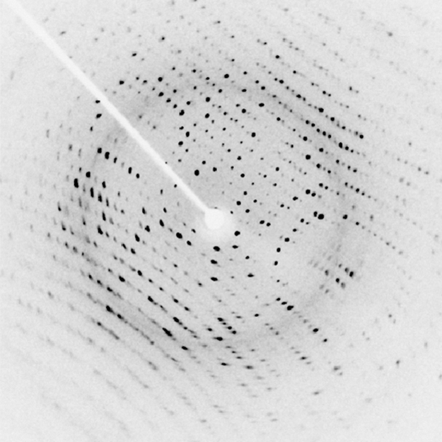
[link] shows a diffraction pattern produced by the scattering of x rays from a crystal. This process is known as x-ray crystallography because of the information it can yield about crystal structure, and it was the type of data Rosalind Franklin supplied to Watson and Crick for DNA. Not only do x rays confirm the size and shape of atoms, they give information on the atomic arrangements in materials. For example, current research in high-temperature superconductors involves complex materials whose lattice arrangements are crucial to obtaining a superconducting material. These can be studied using x-ray crystallography.
Historically, the scattering of x rays from crystals was used to prove that x rays are energetic EM waves. This was suspected from the time of the discovery of x rays in 1895, but it was not until 1912 that the German Max von Laue (1879–1960) convinced two of his colleagues to scatter x rays from crystals. If a diffraction pattern is obtained, he reasoned, then the x rays must be waves, and their wavelength could be determined. (The spacing of atoms in various crystals was reasonably well known at the time, based on good values for Avogadro’s number.) The experiments were convincing, and the 1914 Nobel Prize in Physics was given to von Laue for his suggestion leading to the proof that x rays are EM waves. In 1915, the unique father-and-son team of Sir William Henry Bragg and his son Sir William Lawrence Bragg were awarded a joint Nobel Prize for inventing the x-ray spectrometer and the then-new science of x-ray analysis. The elder Bragg had migrated to Australia from England just after graduating in mathematics. He learned physics and chemistry during his career at the University of Adelaide. The younger Bragg was born in Adelaide but went back to the Cavendish Laboratories in England to a career in x-ray and neutron crystallography; he provided support for Watson, Crick, and Wilkins for their work on unraveling the mysteries of DNA and to Max Perutz for his 1962 Nobel Prize-winning work on the structure of hemoglobin. Here again, we witness the enabling nature of physics—establishing instruments and designing experiments as well as solving mysteries in the biomedical sciences.
Certain other uses for x rays will be studied in later chapters. X rays are useful in the treatment of cancer because of the inhibiting effect they have on cell reproduction. X rays observed coming from outer space are useful in determining the nature of their sources, such as neutron stars and possibly black holes. Created in nuclear bomb explosions, x rays can also be used to detect clandestine atmospheric tests of these weapons. X rays can cause excitations of atoms, which then fluoresce (emitting characteristic EM radiation), making x-ray-induced fluorescence a valuable analytical tool in a range of fields from art to archaeology.
Explain why characteristic x rays are the most energetic in the EM emission spectrum of a given element.
Why does the energy of characteristic x rays become increasingly greater for heavier atoms?
Observers at a safe distance from an atmospheric test of a nuclear bomb feel its heat but receive none of its copious x rays. Why is air opaque to x rays but transparent to infrared?
Lasers are used to burn and read CDs. Explain why a laser that emits blue light would be capable of burning and reading more information than one that emits infrared.
Crystal lattices can be examined with x rays but not UV. Why?
CT scanners do not detect details smaller than about 0.5 mm. Is this limitation due to the wavelength of x rays? Explain.
(a) What is the shortest-wavelength x-ray radiation that can be generated in an x-ray tube with an applied voltage of 50.0 kV? (b) Calculate the photon energy in eV. (c) Explain the relationship of the photon energy to the applied voltage.
(a)
(b) 50.0 keV
(c) The photon energy is simply the applied voltage times the electron charge, so the value of the voltage in volts is the same as the value of the energy in electron volts.
A color television tube also generates some x rays when its electron beam strikes the screen. What is the shortest wavelength of these x rays, if a 30.0-kV potential is used to accelerate the electrons? (Note that TVs have shielding to prevent these x rays from exposing viewers.)
An x ray tube has an applied voltage of 100 kV. (a) What is the most energetic x-ray photon it can produce? Express your answer in electron volts and joules. (b) Find the wavelength of such an X–ray.
(a) ,
(b)
The maximum characteristic x-ray photon energy comes from the capture of a free electron into a shell vacancy. What is this photon energy in keV for tungsten, assuming the free electron has no initial kinetic energy?
What are the approximate energies of the and x rays for copper?
(a) 8.00 keV
(b) 9.48 keV
Notification Switch
Would you like to follow the 'College physics' conversation and receive update notifications?

|
|
|
|
|

|
|

|
|

|
|

|
|

|
|

|
|
|
|
|

|
|

|
|

|