<< Chapter < Page Chapter >> Page >

Development of the embryonic disc

This image shows the development of the amniotic cavity and the location of the embryonic disc.
Formation of the embryonic disc leaves spaces on either side that develop into the amniotic cavity and the yolk sac.

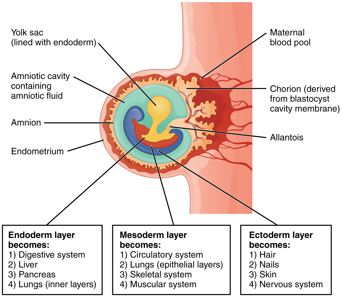
On the ventral side of the embryonic disc, opposite the amnion, cells in the lower layer of the embryonic disk (the hypoblast    ) extend into the blastocyst cavity and form a yolk sac    . The yolk sac supplies some nutrients absorbed from the trophoblast and also provides primitive blood circulation to the developing embryo for the second and third week of development. When the placenta takes over nourishing the embryo at approximately week 4, the yolk sac has been greatly reduced in size and its main function is to serve as the source of blood cells and germ cells (cells that will give rise to gametes). During week 3, a finger-like outpocketing of the yolk sac develops into the allantois    , a primitive excretory duct of the embryo that will become part of the urinary bladder. Together, the stalks of the yolk sac and allantois establish the outer structure of the umbilical cord.

The last of the extra-embryonic membranes is the chorion    , which is the one membrane that surrounds all others. The development of the chorion will be discussed in more detail shortly, as it relates to the growth and development of the placenta.

Embryogenesis

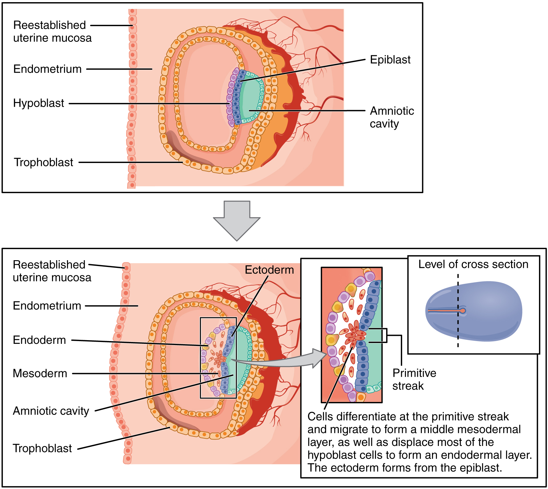
As the third week of development begins, the two-layered disc of cells becomes a three-layered disc through the process of gastrulation    , during which the cells transition from totipotency to multipotency. The embryo, which takes the shape of an oval-shaped disc, forms an indentation called the primitive streak    along the dorsal surface of the epiblast. A node at the caudal or “tail” end of the primitive streak emits growth factors that direct cells to multiply and migrate. Cells migrate toward and through the primitive streak and then move laterally to create two new layers of cells. The first layer is the endoderm    , a sheet of cells that displaces the hypoblast and lies adjacent to the yolk sac. The second layer of cells fills in as the middle layer, or mesoderm    . The cells of the epiblast that remain (not having migrated through the primitive streak) become the ectoderm    ( [link] ).

Germ layers

This image shows the different germ layers. The top panel shows the epiblast and trophoblast cells in the early stages of development. The bottom panel shows the three germ layers: the endoderm, ectoderm, and mesoderm. All the other major parts are also labeled.
Formation of the three primary germ layers occurs during the first 2 weeks of development. The embryo at this stage is only a few millimeters in length.

Each of these germ layers will develop into specific structures in the embryo. Whereas the ectoderm and endoderm form tightly connected epithelial sheets, the mesodermal cells are less organized and exist as a loosely connected cell community. The ectoderm gives rise to cell lineages that differentiate to become the central and peripheral nervous systems, sensory organs, epidermis, hair, and nails. Mesodermal cells ultimately become the skeleton, muscles, connective tissue, heart, blood vessels, and kidneys. The endoderm goes on to form the epithelial lining of the gastrointestinal tract, liver, and pancreas, as well as the lungs ( [link] ).

Fates of germ layers in embryo

This image shows the structure of the embryo in the third week of development. Under the image, three callouts list the different organ systems into which each germ layer develops.
Following gastrulation of the embryo in the third week, embryonic cells of the ectoderm, mesoderm, and endoderm begin to migrate and differentiate into the cell lineages that will give rise to mature organs and organ systems in the infant.

Questions & Answers

how did the oxygen help a human being
Achol Reply
how did the nutrition help the plants
Achol Reply
Biology is a branch of Natural science which deals/About living Organism.
Ahmedin Reply
what is phylogeny
Odigie Reply
evolutionary history and relationship of an organism or group of organisms
AI-Robot
ok
Deng
what is biology
Hajah Reply
cell is the smallest unit of the humanity biologically
Abraham
what is biology
Victoria Reply
what is biology
Abraham
HOW CAN MAN ORGAN FUNCTION
Alfred Reply
the diagram of the digestive system
Assiatu Reply
allimentary cannel
Ogenrwot
How does twins formed
William Reply
They formed in two ways first when one sperm and one egg are splited by mitosis or two sperm and two eggs join together
Oluwatobi
what is genetics
Josephine Reply
Genetics is the study of heredity
Misack
how does twins formed?
Misack
What is manual
Hassan Reply
discuss biological phenomenon and provide pieces of evidence to show that it was responsible for the formation of eukaryotic organelles
Joseph Reply
what is biology
Yousuf Reply
the study of living organisms and their interactions with one another and their environment.
Wine
discuss the biological phenomenon and provide pieces of evidence to show that it was responsible for the formation of eukaryotic organelles in an essay form
Joseph Reply
what is the blood cells
Shaker Reply
list any five characteristics of the blood cells
Shaker
Got questions? Join the online conversation and get instant answers!
Jobilize.com Reply

Get Jobilize Job Search Mobile App in your pocket Now!

Get it on Google Play Download on the App Store Now


Source:  OpenStax, Genetics and evolution. OpenStax CNX. Aug 07, 2014 Download for free at https://legacy.cnx.org/content/col11595/1.2
Google Play and the Google Play logo are trademarks of Google Inc.

Notification Switch

Would you like to follow the 'Genetics and evolution' conversation and receive update notifications?

Ask