<< Chapter < Page Chapter >> Page >
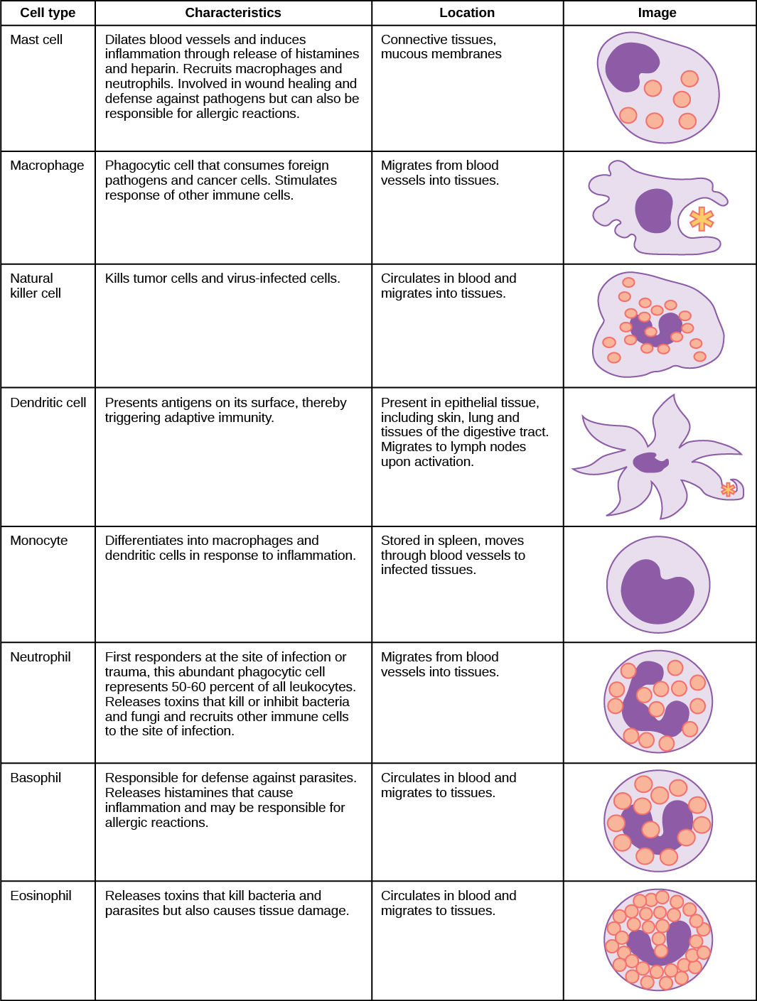
 Table shows various types of white blood cells and describes their function. Mast cells, natural killer cells, neutrophils, basophils and eosinophils are all filled with granules and have a horseshoe-shaped nucleus. Macrophages are irregular in shape, with a round nucleus. Dendrites have star-like projections and a small horseshoe shaped nucleus. Mast cells dilate blood vessels and induce inflammation through release of histamines and heparin. They also recruit macrophages and neutrophils, and are involved in wound healing and defense against pathogens, but can also be responsible for allergic reactions. They are found in connective tissue and mucous membranes. Macrophages are phagocytic cells that consume foreign pathogens and cancer cells. They stimulate response of other immune cells and migrate from blood vessels into tissues. Natural killer cells kill tumor cells and virus-infected cells. They circulate in blood and migrate into tissues. Dendritic cells present antigens on their surface, thereby triggering adaptive immunity. They are present in tissues in epithelial tissue, including skin, lung and tissues of the digestive tract. Migrate to lymph nodes upon activation. Monocytes differentiate into macrophages and dendritic cells in response to inflammation. They are stored in spleen, move through blood vessels to infected tissues. Neutrophils are first responders at the site of infection or trauma, these abundant phagocytic cell representing 50-60% of all leukocytes. Release toxins that kill or inhibit bacteria and fungi and recruit other immune cells to the site of infection. They migrate from blood vessels into tissues. Basophils are responsible for defense against parasites. They release histamines that cause inflammation and may be responsible for allergic reactions. They circulate in blood and migrate to tissues. Eosinophils release toxins that kill bacteria and parasites but also causes tissue damage. They circulate in blood and migrate to tissues.
The characteristics and location of cells involved in the innate immune system are described. (credit: modification of work by NIH)
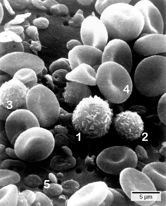
Micrograph shows a blood smear. Red blood cells are disk-shaped, and pinched together in the center. Monocytes, lymphocytes and neutrophils are all ball-shaped and fuzzy. Platelets are small, flat disks.
Cells of the blood include (1) monocytes, (2) lymphocytes, (3) neutrophils, (4) red blood cells, and (5) platelets. Note the very similar morphologies of the leukocytes (1, 2, 3). (credit: modification of work by Bruce Wetzel, Harry Schaefer, NCI; scale-bar data from Matt Russell)

Cytokine release affect

The binding of PRRs with PAMPs triggers the release of cytokines, which signal that a pathogen is present and needs to be destroyed along with any infected cells. A cytokine    is a chemical messenger that regulates cell differentiation (form and function), proliferation (production), and gene expression to affect immune responses. At least 40 types of cytokines exist in humans that differ in terms of the cell type that produces them, the cell type that responds to them, and the changes they produce. One type cytokine, interferon, is illustrated in [link] .

One subclass of cytokines is the interleukin (IL), so named because they mediate interactions between leukocytes (white blood cells). Interleukins are involved in bridging the innate and adaptive immune responses. In addition to being released from cells after PAMP recognition, cytokines are released by the infected cells which bind to nearby uninfected cells and induce those cells to release cytokines, which results in a cytokine burst.

A second class of early-acting cytokines is interferons, which are released by infected cells as a warning to nearby uninfected cells. One of the functions of an interferon    is to inhibit viral replication. They also have other important functions, such as tumor surveillance. Interferons work by signaling neighboring uninfected cells to destroy RNA and reduce protein synthesis, signaling neighboring infected cells to undergo apoptosis (programmed cell death), and activating immune cells.

In response to interferons, uninfected cells alter their gene expression, which increases the cells’ resistance to infection. One effect of interferon-induced gene expression is a sharply reduced cellular protein synthesis. Virally infected cells produce more viruses by synthesizing large quantities of viral proteins. Thus, by reducing protein synthesis, a cell becomes resistant to viral infection.
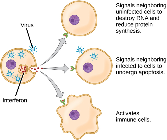
Illustration shows a virus-infected cell secreting interferon, which binds to receptors of neighboring cells. Interferon signals neighboring uninfected cells to destroy RNA and reduce protein synthesis, thus making it more difficult for virus to infect the cell. It signals neighboring infected cells to undergo apoptosis. It also activates nearby immune cells.
Interferons are cytokines that are released by a cell infected with a virus. Response of neighboring cells to interferon helps stem the infection.

Phagocytosis and inflammation

The first cytokines to be produced are pro-inflammatory; that is, they encourage inflammation    , the localized redness, swelling, heat, and pain that result from the movement of leukocytes and fluid through increasingly permeable capillaries to a site of infection. The population of leukocytes that arrives at an infection site depends on the nature of the infecting pathogen. Both macrophages and dendritic cells engulf pathogens and cellular debris through phagocytosis. A neutrophil    is also a phagocytic leukocyte that engulfs and digests pathogens. Neutrophils, shown in [link] , are the most abundant leukocytes of the immune system. Neutrophils have a nucleus with two to five lobes, and they contain organelles, called lysosomes, that digest engulfed pathogens. An eosinophil    is a leukocyte that works with other eosinophils to surround a parasite; it is involved in the allergic response and in protection against helminthes (parasitic worms).

Get Jobilize Job Search Mobile App in your pocket Now!

Get it on Google Play Download on the App Store Now


Source:  OpenStax, Biology. OpenStax CNX. Feb 29, 2016 Download for free at http://cnx.org/content/col11448/1.10
Google Play and the Google Play logo are trademarks of Google Inc.

Notification Switch

Would you like to follow the 'Biology' conversation and receive update notifications?

Ask