<< Chapter < Page Chapter >> Page >

Modes of glandular secretion

These three diagrams show the three modes of secretion. All three diagrams show three orange cells in a line with attached to a basement membrane. Each cell has a large nucleus in its lower half. The upper half of each cell contains a Golgi apparatus, which appears like an upside down jellyfish. Yellow secretory vesicles are budding from the top end of the Golgi apparatus. Each vesicle contains several orange circles, which are the secreted substance. In merocrine secretion, the secretory vesicles travel to the top edge of the cells and release the secretion from the cell by melding with the cell membrane. In apocrine secretion, the top third of the cell, which contains the secretory vesicles, pinches in at the sides and then completely disconnects above the Golgi complex. The pinched off portion of the cell is the secretion, as it contains the majority of the secretory vesicles. In holocrine secretion, the upper third of the cell, just above the Golgi complex, forms many finger like projections. Each projection contains several vesicles. The tips of the projections that contain secretory vesicles bud off from the cell. In this method of secretion, the mature cell eventually dies and becomes the secretory product.
(a) In merocrine secretion, the cell remains intact. (b) In apocrine secretion, the apical portion of the cell is released, as well. (c) In holocrine secretion, the cell is destroyed as it releases its product and the cell itself becomes part of the secretion.

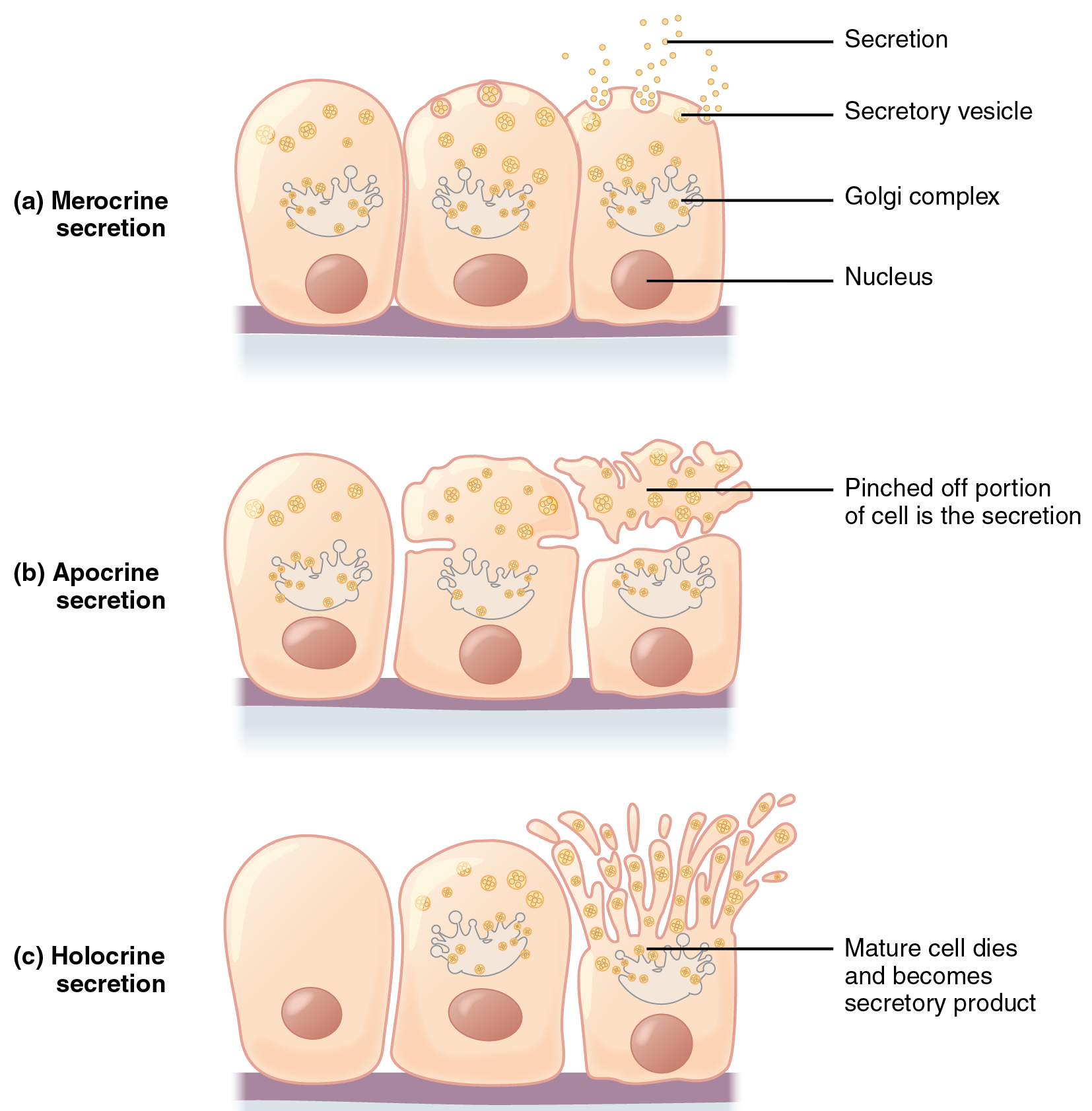
Apocrine secretion accumulates near the apical portion of the cell. That portion of the cell and its secretory contents pinch off from the cell and are released. The sweat glands of the armpit are classified as apocrine glands. Both merocrine and apocrine glands continue to produce and secrete their contents with little damage caused to the cell because the nucleus and golgi regions remain intact after secretion.

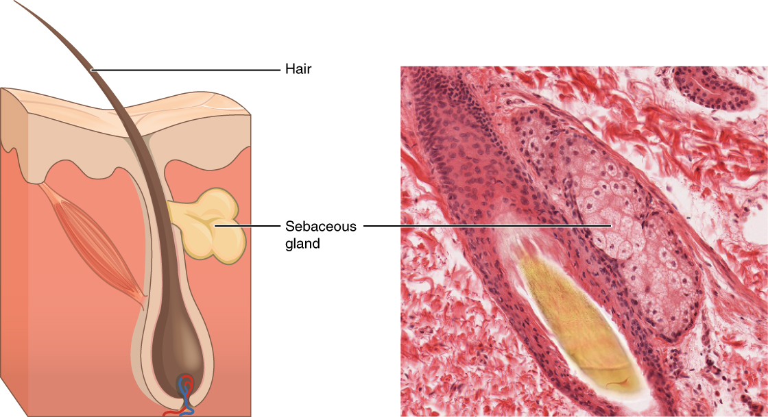
In contrast, the process of holocrine secretion    involves the rupture and destruction of the entire gland cell. The cell accumulates its secretory products and releases them only when it bursts. New gland cells differentiate from cells in the surrounding tissue to replace those lost by secretion. The sebaceous glands that produce the oils on the skin and hair are holocrine glands/cells ( [link] ).

Sebaceous glands

Image A depicts a cross section of the skin layers. The surface of the skin is at the top of the diagram, with the outer layer occupying about one fifth of the cross section. The outer layer has an irregular border with the inner skin layer, which occupies the remainder of the cross section. A hair follicle is embedded within the inner layer. However, the outer layer actually invaginates into the inner layer around the outside of the follicle, completely sheathing the follicle. The follicle has a bulb at its bottom that is connected to blood vessels. The hair projects from the bulb and travels through the sheath to erupt from the skin surface. The sebaceous gland is an irregular, yellow structure attached at the midpoint of the hair shaft near the border between the inner and outer layers of skin. Its duct actually connects into the side of the hair follicle. Image B shows a micrograph of a sebaceous gland connected to a hair follicle. The bulb of the hair follicle is evident in the micrograph as a bundle of cell surrounding the growing hair at its center. The sebaceous gland is connected to the right of the follicle bulb. The gland appears as an oval shaped mass of pink staining, cube shaped cells with purple nuclei.
These glands secrete oils that lubricate and protect the skin. They are holocrine glands and they are destroyed after releasing their contents. New glandular cells form to replace the cells that are lost. LM × 400. (Micrograph provided by the Regents of University of Michigan Medical School © 2012)

Glands are also named after the products they produce. The serous gland    produces watery, blood-plasma-like secretions rich in enzymes such as alpha amylase, whereas the mucous gland    releases watery to viscous products rich in the glycoprotein mucin. Both serous and mucous glands are common in the salivary glands of the mouth. Mixed exocrine glands contain both serous and mucous glands and release both types of secretions.

Chapter review

In epithelial tissue, cells are closely packed with little or no extracellular matrix except for the basal lamina that separates the epithelium from underlying tissue. The main functions of epithelia are protection from the environment, coverage, secretion and excretion, absorption, and filtration. Cells are bound together by tight junctions that form an impermeable barrier. They can also be connected by gap junctions, which allow free exchange of soluble molecules between cells, and anchoring junctions, which attach cell to cell or cell to matrix. The different types of epithelial tissues are characterized by their cellular shapes and arrangements: squamous, cuboidal, or columnar epithelia. Single cell layers form simple epithelia, whereas stacked cells form stratified epithelia. Very few capillaries penetrate these tissues.

Glands are secretory tissues and organs that are derived from epithelial tissues. Exocrine glands release their products through ducts. Endocrine glands secrete hormones directly into the interstitial fluid and blood stream. Glands are classified both according to the type of secretion and by their structure. Merocrine glands secrete products as they are synthesized. Apocrine glands release secretions by pinching off the apical portion of the cell, whereas holocrine gland cells store their secretions until they rupture and release their contents. In this case, the cell becomes part of the secretion.

Watch this video to find out more about the anatomy of epithelial tissues. Where in the body would one find non-keratinizing stratified squamous epithelium?

The inside of the mouth, esophagus, vaginal canal, and anus.

Got questions? Get instant answers now!

Questions & Answers

how does Neisseria cause meningitis
Nyibol Reply
what is microbiologist
Muhammad Reply
what is errata
Muhammad
is the branch of biology that deals with the study of microorganisms.
Ntefuni Reply
What is microbiology
Mercy Reply
studies of microbes
Louisiaste
when we takee the specimen which lumbar,spin,
Ziyad Reply
How bacteria create energy to survive?
Muhamad Reply
Bacteria doesn't produce energy they are dependent upon their substrate in case of lack of nutrients they are able to make spores which helps them to sustain in harsh environments
_Adnan
But not all bacteria make spores, l mean Eukaryotic cells have Mitochondria which acts as powerhouse for them, since bacteria don't have it, what is the substitution for it?
Muhamad
they make spores
Louisiaste
what is sporadic nd endemic, epidemic
Aminu Reply
the significance of food webs for disease transmission
Abreham
food webs brings about an infection as an individual depends on number of diseased foods or carriers dully.
Mark
explain assimilatory nitrate reduction
Esinniobiwa Reply
Assimilatory nitrate reduction is a process that occurs in some microorganisms, such as bacteria and archaea, in which nitrate (NO3-) is reduced to nitrite (NO2-), and then further reduced to ammonia (NH3).
Elkana
This process is called assimilatory nitrate reduction because the nitrogen that is produced is incorporated in the cells of microorganisms where it can be used in the synthesis of amino acids and other nitrogen products
Elkana
Examples of thermophilic organisms
Shu Reply
Give Examples of thermophilic organisms
Shu
advantages of normal Flora to the host
Micheal Reply
Prevent foreign microbes to the host
Abubakar
they provide healthier benefits to their hosts
ayesha
They are friends to host only when Host immune system is strong and become enemies when the host immune system is weakened . very bad relationship!
Mark
what is cell
faisal Reply
cell is the smallest unit of life
Fauziya
cell is the smallest unit of life
Akanni
ok
Innocent
cell is the structural and functional unit of life
Hasan
is the fundamental units of Life
Musa
what are emergency diseases
Micheal Reply
There are nothing like emergency disease but there are some common medical emergency which can occur simultaneously like Bleeding,heart attack,Breathing difficulties,severe pain heart stock.Hope you will get my point .Have a nice day ❣️
_Adnan
define infection ,prevention and control
Innocent
I think infection prevention and control is the avoidance of all things we do that gives out break of infections and promotion of health practices that promote life
Lubega
Heyy Lubega hussein where are u from?
_Adnan
en français
Adama
which site have a normal flora
ESTHER Reply
Many sites of the body have it Skin Nasal cavity Oral cavity Gastro intestinal tract
Safaa
skin
Asiina
skin,Oral,Nasal,GIt
Sadik
How can Commensal can Bacteria change into pathogen?
Sadik
How can Commensal Bacteria change into pathogen?
Sadik
all
Tesfaye
by fussion
Asiina
what are the advantages of normal Flora to the host
Micheal
what are the ways of control and prevention of nosocomial infection in the hospital
Micheal
what is inflammation
Shelly Reply
part of a tissue or an organ being wounded or bruised.
Wilfred
what term is used to name and classify microorganisms?
Micheal Reply
Binomial nomenclature
adeolu
Got questions? Join the online conversation and get instant answers!
Jobilize.com Reply

Get Jobilize Job Search Mobile App in your pocket Now!

Get it on Google Play Download on the App Store Now


Source:  OpenStax, Anatomy & Physiology. OpenStax CNX. Feb 04, 2016 Download for free at http://legacy.cnx.org/content/col11496/1.8
Google Play and the Google Play logo are trademarks of Google Inc.

Notification Switch

Would you like to follow the 'Anatomy & Physiology' conversation and receive update notifications?

Ask