<< Chapter < Page Chapter >> Page >

Planes of the body

This illustration shows a female viewed from her right, front side. The anatomical planes are depicted as blue rectangles passing through the woman’s body. The frontal or coronal plane enters through the right side of the body, passes through the body, and exits from the left side. It divides the body into front (anterior) and back (posterior) halves. The sagittal plane enters through the back and emerges through the front of the body. It divides the body into right and left halves. The transverse plane passes through the body perpendicular to the frontal and sagittal planes. This plane is a cross section which divides the body into upper and lower halves.
The three planes most commonly used in anatomical and medical imaging are the sagittal, frontal (or coronal), and transverse plane.

Body cavities and serous membranes

The body maintains its internal organization by means of membranes, sheaths, and other structures that separate compartments. The dorsal (posterior) cavity and the ventral (anterior) cavity are the largest body compartments ( [link] ). These cavities contain and protect delicate internal organs, and the ventral cavity allows for significant changes in the size and shape of the organs as they perform their functions. The lungs, heart, stomach, and intestines, for example, can expand and contract without distorting other tissues or disrupting the activity of nearby organs.

Dorsal and ventral body cavities

This illustration shows a lateral and anterior view of the body and highlights the body cavities with different colors. The cranial cavity is a large, bean-shaped cavity filling most of the upper skull where the brain is located. The vertebral cavity is a very narrow, thread-like cavity running from the cranial cavity down the entire length of the spinal cord. Together the cranial cavity and vertebral cavity can be referred to as the dorsal body cavity. The thoracic cavity consists of three cavities that fill the interior area of the chest. The two pleural cavities are situated on both sides of the body, anterior to the spine and lateral to the breastbone. The superior mediastinum is a wedge-shaped cavity located between the superior regions of the two thoracic cavities. The pericardial cavity within the mediastinum is located at the center of the chest below the superior mediastinum. The pericardial cavity roughly outlines the shape of the heart. The diaphragm divides the thoracic and the abdominal cavities. The abdominal cavity occupies the entire lower half of the trunk, anterior to the spine. Just under the abdominal cavity, anterior to the buttocks, is the pelvic cavity. The pelvic cavity is funnel shaped and is located inferior and anterior to the abdominal cavity. Together the abdominal and pelvic cavity can be referred to as the abdominopelvic cavity while the thoracic, abdominal, and pelvic cavities together can be referred to as the ventral body cavity.
The ventral cavity includes the thoracic and abdominopelvic cavities and their subdivisions. The dorsal cavity includes the cranial and spinal cavities.

Subdivisions of the posterior (dorsal) and anterior (ventral) cavities

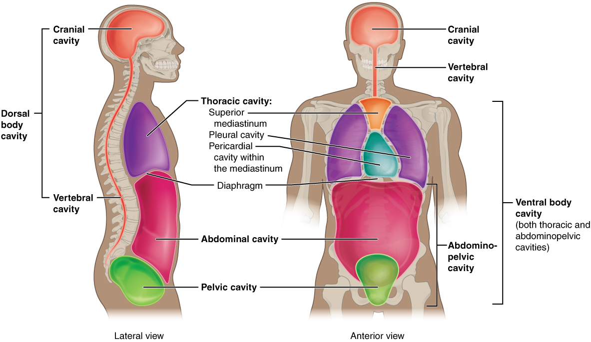
The posterior (dorsal) and anterior (ventral) cavities are each subdivided into smaller cavities. In the posterior (dorsal) cavity, the cranial cavity    houses the brain, and the spinal cavity    (or vertebral cavity) encloses the spinal cord. Just as the brain and spinal cord make up a continuous, uninterrupted structure, the cranial and spinal cavities that house them are also continuous. The brain and spinal cord are protected by the bones of the skull and vertebral column and by cerebrospinal fluid, a colorless fluid produced by the brain, which cushions the brain and spinal cord within the posterior (dorsal) cavity.

The anterior (ventral) cavity has two main subdivisions: the thoracic cavity and the abdominopelvic cavity (see [link] ). The thoracic cavity    is the more superior subdivision of the anterior cavity, and it is enclosed by the rib cage. The thoracic cavity contains the lungs and the heart, which is located in the mediastinum. The diaphragm forms the floor of the thoracic cavity and separates it from the more inferior abdominopelvic cavity. The abdominopelvic cavity    is the largest cavity in the body. Although no membrane physically divides the abdominopelvic cavity, it can be useful to distinguish between the abdominal cavity, the division that houses the digestive organs, and the pelvic cavity, the division that houses the organs of reproduction.

Abdominal regions and quadrants

To promote clear communication, for instance about the location of a patient’s abdominal pain or a suspicious mass, health care providers typically divide up the cavity into either nine regions or four quadrants ( [link] ).

Regions and quadrants of the peritoneal cavity

This illustration has two parts. Part A shows the abdominopelvic regions. These regions divide the abdomen into nine squares. The upper right square is the right hypochondriac region and contains the base of the right ribs. The upper left square is the left hypochondriac region and contains the base of the left ribs. The epigastric region is the upper central square and contains the bottom edge of the liver as well as the upper areas of the stomach. The diaphragm curves like an upside down U over these three regions. The central right region is called the right lumbar region and contains the ascending colon and the right edge of the small intestines. The central square contains the transverse colon and the upper regions of the small intestines. The left lumbar region contains the left edge of the transverse colon and the left edge of the small intestine. The lower right square is the right iliac region and contains the right pelvic bones and the ascending colon. The lower left square is the left iliac region and contains the left pelvic bone and the lower left regions of the small intestine. The lower central square contains the bottom of the pubic bones, upper regions of the bladder and the lower region of the small intestine. Part B shows four abdominopelvic quadrants. The right upper quadrant (RUQ) includes the lower right ribs, right side of the liver, and right side of the transverse colon. The left upper quadrant (LUQ) includes the lower left ribs, stomach, and upper left area of the transverse colon. The right lower quadrant (RLQ) includes the right half of the small intestines, ascending colon, right pelvic bone and upper right area of the bladder. The left lower quadrant (LLQ) contains the left half of the small intestine and left pelvic bone.
There are (a) nine abdominal regions and (b) four abdominal quadrants in the peritoneal cavity.

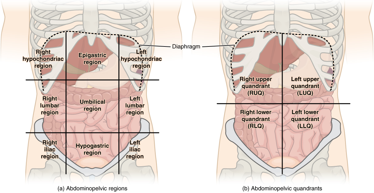
The more detailed regional approach subdivides the cavity with one horizontal line immediately inferior to the ribs and one immediately superior to the pelvis, and two vertical lines drawn as if dropped from the midpoint of each clavicle (collarbone). There are nine resulting regions. The simpler quadrants approach, which is more commonly used in medicine, subdivides the cavity with one horizontal and one vertical line that intersect at the patient’s umbilicus (navel).

Membranes of the anterior (ventral) body cavity

A serous membrane    (also referred to a serosa) is one of the thin membranes that cover the walls and organs in the thoracic and abdominopelvic cavities. The parietal layers of the membranes line the walls of the body cavity (pariet- refers to a cavity wall). The visceral layer of the membrane covers the organs (the viscera). Between the parietal and visceral layers is a very thin, fluid-filled serous space, or cavity ( [link] ).

Serous membrane

This diagram shows the pericardium on the left next to an analogy of a hand punching a balloon on the right. The pericardium is a two-layered sac that surrounds the entire heart except where the blood vessels emerge on the heart’s superior side. The pericardium has two layers because it folds over itself in the shape of the letter U. The inner layer that borders the heart is the visceral pericardium while the outer layer is the parietal pericardium. The space between the two layers is called the pericardial cavity. The heart sits in the cavity much like a fist punching into a balloon. The balloon surrounds the lower part of the fist with a two-layered sac, with the top of the balloon, where it contacts the fist, being analogous to the visceral pericardium. The bottom of the balloon, where it is tied off, is analogous to the parietal pericardium. The air within the balloon is analogous to the pericardial cavity.
Serous membrane lines the pericardial cavity and reflects back to cover the heart—much the same way that an underinflated balloon would form two layers surrounding a fist.

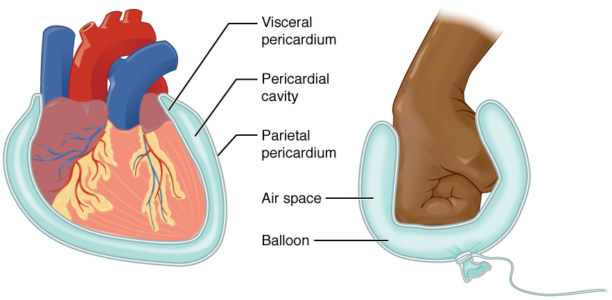
There are three serous cavities and their associated membranes. The pleura    is the serous membrane that surrounds the lungs in the pleural cavity; the pericardium    is the serous membrane that surrounds the heart in the pericardial cavity; and the peritoneum    is the serous membrane that surrounds several organs in the abdominopelvic cavity.The serous membranes form fluid-filled sacs, or cavities, that are meant to cushion and reduce friction on internal organs when they move, such as when the lungs inflate or the heart beats. Both the parietal and visceral serosa secrete the thin, slippery serous fluid located within the serous cavities. The pleural cavity reduces friction between the lungs and the body wall. Likewise, the pericardial cavity reduces friction between the heart and the wall of the pericardium. The peritoneal cavity reduces friction between the abdominal and pelvic organs and the body wall. Therefore, serous membranes provide additional protection to the viscera they enclose by reducing friction that could lead to inflammation of the organs.

Chapter review

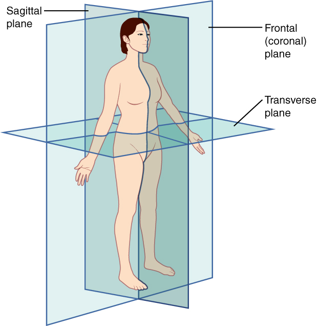
Ancient Greek and Latin words are used to build anatomical terms. A standard reference position for mapping the body’s structures is the normal anatomical position. Regions of the body are identified using terms such as “occipital” that are more precise than common words and phrases such as “the back of the head.” Directional terms such as anterior and posterior are essential for accurately describing the relative locations of body structures. Images of the body’s interior commonly align along one of three planes: the sagittal, frontal, or transverse. The body’s organs are organized in one of two main cavities—dorsal (also referred to posterior) and ventral (also referred to anterior)—which are further sub-divided according to the structures present in each area. The serous membranes have two layers—parietal and visceral—surrounding a fluid filled space. Serous membranes cover the lungs (pleural serosa), heart (pericardial serosa), and some abdominopelvic organs (peritoneal serosa).

Questions & Answers

A golfer on a fairway is 70 m away from the green, which sits below the level of the fairway by 20 m. If the golfer hits the ball at an angle of 40° with an initial speed of 20 m/s, how close to the green does she come?
Aislinn Reply
cm
tijani
what is titration
John Reply
what is physics
Siyaka Reply
A mouse of mass 200 g falls 100 m down a vertical mine shaft and lands at the bottom with a speed of 8.0 m/s. During its fall, how much work is done on the mouse by air resistance
Jude Reply
Can you compute that for me. Ty
Jude
what is the dimension formula of energy?
David Reply
what is viscosity?
David
what is inorganic
emma Reply
what is chemistry
Youesf Reply
what is inorganic
emma
Chemistry is a branch of science that deals with the study of matter,it composition,it structure and the changes it undergoes
Adjei
please, I'm a physics student and I need help in physics
Adjanou
chemistry could also be understood like the sexual attraction/repulsion of the male and female elements. the reaction varies depending on the energy differences of each given gender. + masculine -female.
Pedro
A ball is thrown straight up.it passes a 2.0m high window 7.50 m off the ground on it path up and takes 1.30 s to go past the window.what was the ball initial velocity
Krampah Reply
2. A sled plus passenger with total mass 50 kg is pulled 20 m across the snow (0.20) at constant velocity by a force directed 25° above the horizontal. Calculate (a) the work of the applied force, (b) the work of friction, and (c) the total work.
Sahid Reply
you have been hired as an espert witness in a court case involving an automobile accident. the accident involved car A of mass 1500kg which crashed into stationary car B of mass 1100kg. the driver of car A applied his brakes 15 m before he skidded and crashed into car B. after the collision, car A s
Samuel Reply
can someone explain to me, an ignorant high school student, why the trend of the graph doesn't follow the fact that the higher frequency a sound wave is, the more power it is, hence, making me think the phons output would follow this general trend?
Joseph Reply
Nevermind i just realied that the graph is the phons output for a person with normal hearing and not just the phons output of the sound waves power, I should read the entire thing next time
Joseph
Follow up question, does anyone know where I can find a graph that accuretly depicts the actual relative "power" output of sound over its frequency instead of just humans hearing
Joseph
"Generation of electrical energy from sound energy | IEEE Conference Publication | IEEE Xplore" ***ieeexplore.ieee.org/document/7150687?reload=true
Ryan
what's motion
Maurice Reply
what are the types of wave
Maurice
answer
Magreth
progressive wave
Magreth
hello friend how are you
Muhammad Reply
fine, how about you?
Mohammed
hi
Mujahid
A string is 3.00 m long with a mass of 5.00 g. The string is held taut with a tension of 500.00 N applied to the string. A pulse is sent down the string. How long does it take the pulse to travel the 3.00 m of the string?
yasuo Reply
Who can show me the full solution in this problem?
Reofrir Reply
Got questions? Join the online conversation and get instant answers!
Jobilize.com Reply

Get Jobilize Job Search Mobile App in your pocket Now!

Get it on Google Play Download on the App Store Now


Source:  OpenStax, Anatomy & Physiology. OpenStax CNX. Feb 04, 2016 Download for free at http://legacy.cnx.org/content/col11496/1.8
Google Play and the Google Play logo are trademarks of Google Inc.

Notification Switch

Would you like to follow the 'Anatomy & Physiology' conversation and receive update notifications?

Ask