<< Chapter < Page Chapter >> Page >
By the end of this section, you will be able to:
  • Identify the anatomical features of a bone
  • Define and list examples of bone markings
  • Describe the histology of bone tissue
  • Compare and contrast compact and spongy bone
  • Identify the structures that compose compact and spongy bone
  • Describe how bones are nourished and innervated

Bone tissue (osseous tissue) differs greatly from other tissues in the body. Bone is hard and many of its functions depend on that characteristic hardness. Later discussions in this chapter will show that bone is also dynamic in that its shape adjusts to accommodate stresses. This section will examine the gross anatomy of bone first and then move on to its histology.

Gross anatomy of bone

The structure of a long bone allows for the best visualization of all of the parts of a bone ( [link] ). A long bone has two parts: the diaphysis    and the epiphysis    . The diaphysis is the tubular shaft that runs between the proximal and distal ends of the bone. The hollow region in the diaphysis is called the medullary cavity    , which is filled with yellow marrow. The walls of the diaphysis are composed of dense and hard compact bone    .

Anatomy of a long bone

This illustration depicts an anterior view of the right femur, or thigh bone. The inferior end that connects to the knee is at the bottom of the diagram and the superior end that connects to the hip is at the top of the diagram. The bottom end of the bone contains a smaller lateral bulge and a larger medial bulge. A blue articular cartilage covers the inner half of each bulge as well as the small trench that runs between the bulges. This area of the inferior end of the bone is labeled the distal epiphysis. Above the distal epiphysis is the metaphysis, where the bone tapers from the wide epiphysis into the relatively thin shaft. The entire length of the shaft is the diaphysis. The superior half of the femur is cut away to show its internal contents. The bone is covered with an outer translucent sheet called the periosteum. At the midpoint of the diaphysis, a nutrient artery travels through the periosteum and into the inner layers of the bone. The periosteum surrounds a white cylinder of solid bone labeled compact bone. The cavity at the center of the compact bone is called the medullary cavity. The inner layer of the compact bone that lines the medullary cavity is called the endosteum. Within the diaphysis, the medullary cavity contains a cylinder of yellow bone marrow that is penetrated by the nutrient artery. The superior end of the femur is also connected to the diaphysis by a metaphysis. In this upper metaphysis, the bone gradually widens between the diaphysis and the proximal epiphysis. The proximal epiphysis of the femur is roughly hexagonal in shape. However, the upper right side of the hexagon has a large, protruding knob. The femur connects and rotates within the hip socket at this knob. The knob is covered with a blue colored articular cartilage. The internal anatomy of the upper metaphysis and proximal epiphysis are revealed. The medullary cavity in these regions is filled with the mesh like spongy bone. Red bone marrow occupies the many cavities within the spongy bone. There is a clear, white line separating the spongy bone of the upper metaphysis with that of the proximal epiphysis. This line is labeled the epiphyseal line.
A typical long bone shows the gross anatomical characteristics of bone.

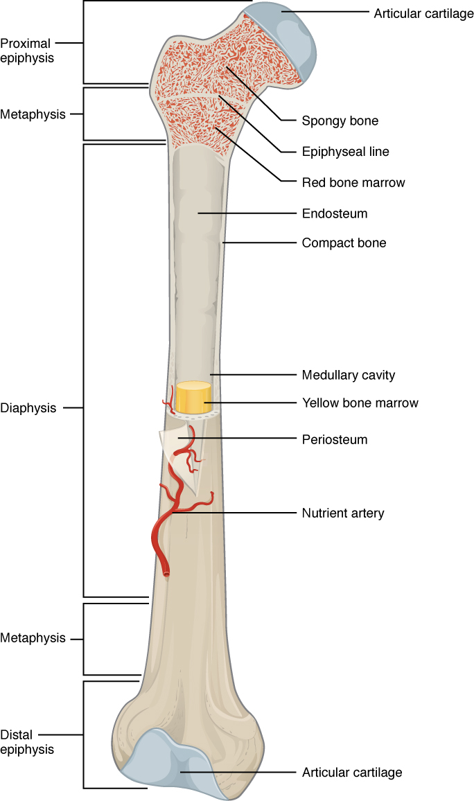
The wider section at each end of the bone is called the epiphysis (plural = epiphyses), which is filled with spongy bone. Red marrow fills the spaces in the spongy bone. Each epiphysis meets the diaphysis at the metaphysis, the narrow area that contains the epiphyseal plate    (growth plate), a layer of hyaline (transparent) cartilage in a growing bone. When the bone stops growing in early adulthood (approximately 18–21 years), the cartilage is replaced by osseous tissue and the epiphyseal plate becomes an epiphyseal line.

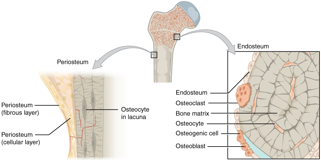
The medullary cavity has a delicate membranous lining called the endosteum    (end- = “inside”; oste- = “bone”), where bone growth, repair, and remodeling occur. The outer surface of the bone is covered with a fibrous membrane called the periosteum    (peri - = “around” or “surrounding”). The periosteum contains blood vessels, nerves, and lymphatic vessels that nourish compact bone. Tendons and ligaments also attach to bones at the periosteum. The periosteum covers the entire outer surface except where the epiphyses meet other bones to form joints ( [link] ). In this region, the epiphyses are covered with articular cartilage    , a thin layer of cartilage that reduces friction and acts as a shock absorber.

Periosteum and endosteum

The top of this illustration shows an anterior view of the proximal end of the femur. The top image has two zoom in boxes. The left box is situated on the border between the diaphysis and the metaphysis. Its callout magnifies the periosteum on the right side of the femur. The view shows that the periosteum contains an outer fibrous layer composed of yellow fibers. The inner layer of the periosteum is called the cellular layer, which is composed of irregularly shaped cells. The cellular layer gradually shrinks in width as it transitions from the metaphysis to the diaphysis. A small blood vessel runs through both layers and enters the bone. The right zoom in box magnifies the endosteum on the left side of the bone. The box is situated just inferior to the border between the diaphysis and the metaphysic. It calls out the inner edge of the compact bone layer. The magnified view shows concentric circles of dark colored bone matrix. Between the circles are small cavities containing orange, diamond-shaped cells labeled osteocytes. The left edge of the bone matrix is lined with a single layer of flattened cells called the endosteum. There is a large cell, labeled an osteoclast, between two of the endosteum cells. The osteoclast is cutting a depression into the bony matrix under the endosteum. At another part of the endosteum, three smaller osteoblasts are secreting a blue substance that builds up the outermost layer of the bony matrix.
The periosteum forms the outer surface of bone, and the endosteum lines the medullary cavity.

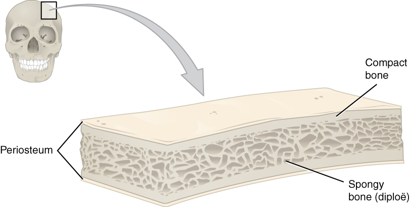
Flat bones, like those of the cranium, consist of a layer of diploë    (spongy bone), lined on either side by a layer of compact bone ( [link] ). The two layers of compact bone and the interior spongy bone work together to protect the internal organs. If the outer layer of a cranial bone fractures, the brain is still protected by the intact inner layer.

Anatomy of a flat bone

This illustration shows a cross section of a cranial bone, constructed somewhat like a sandwich. The topmost and bottommost layers are the thin, translucent, periosteum. The upper and lower periosteum cover an upper and lower layer of compact bone, respectively. The compact bone is solid, with each layer occupying about one tenth of the thickness of the cranial bone. The majority of the cross section is occupied by the spongy bone, or diploe, sandwiched between the upper and lower compact bone. The spongy bone contains many crisscrossing threads of bone. Dark air spaces occur between the threads, giving the bone a porous appearance, much like that of a sponge or Swiss cheese.
This cross-section of a flat bone shows the spongy bone (diploë) lined on either side by a layer of compact bone.

Questions & Answers

Three charges q_{1}=+3\mu C, q_{2}=+6\mu C and q_{3}=+8\mu C are located at (2,0)m (0,0)m and (0,3) coordinates respectively. Find the magnitude and direction acted upon q_{2} by the two other charges.Draw the correct graphical illustration of the problem above showing the direction of all forces.
Kate Reply
To solve this problem, we need to first find the net force acting on charge q_{2}. The magnitude of the force exerted by q_{1} on q_{2} is given by F=\frac{kq_{1}q_{2}}{r^{2}} where k is the Coulomb constant, q_{1} and q_{2} are the charges of the particles, and r is the distance between them.
Muhammed
What is the direction and net electric force on q_{1}= 5µC located at (0,4)r due to charges q_{2}=7mu located at (0,0)m and q_{3}=3\mu C located at (4,0)m?
Kate Reply
what is the change in momentum of a body?
Eunice Reply
what is a capacitor?
Raymond Reply
Capacitor is a separation of opposite charges using an insulator of very small dimension between them. Capacitor is used for allowing an AC (alternating current) to pass while a DC (direct current) is blocked.
Gautam
A motor travelling at 72km/m on sighting a stop sign applying the breaks such that under constant deaccelerate in the meters of 50 metres what is the magnitude of the accelerate
Maria Reply
please solve
Sharon
8m/s²
Aishat
What is Thermodynamics
Muordit
velocity can be 72 km/h in question. 72 km/h=20 m/s, v^2=2.a.x , 20^2=2.a.50, a=4 m/s^2.
Mehmet
A boat travels due east at a speed of 40meter per seconds across a river flowing due south at 30meter per seconds. what is the resultant speed of the boat
Saheed Reply
50 m/s due south east
Someone
which has a higher temperature, 1cup of boiling water or 1teapot of boiling water which can transfer more heat 1cup of boiling water or 1 teapot of boiling water explain your . answer
Ramon Reply
I believe temperature being an intensive property does not change for any amount of boiling water whereas heat being an extensive property changes with amount/size of the system.
Someone
Scratch that
Someone
temperature for any amount of water to boil at ntp is 100⁰C (it is a state function and and intensive property) and it depends both will give same amount of heat because the surface available for heat transfer is greater in case of the kettle as well as the heat stored in it but if you talk.....
Someone
about the amount of heat stored in the system then in that case since the mass of water in the kettle is greater so more energy is required to raise the temperature b/c more molecules of water are present in the kettle
Someone
definitely of physics
Haryormhidey Reply
how many start and codon
Esrael Reply
what is field
Felix Reply
physics, biology and chemistry this is my Field
ALIYU
field is a region of space under the influence of some physical properties
Collete
what is ogarnic chemistry
WISDOM Reply
determine the slope giving that 3y+ 2x-14=0
WISDOM
Another formula for Acceleration
Belty Reply
a=v/t. a=f/m a
IHUMA
innocent
Adah
pratica A on solution of hydro chloric acid,B is a solution containing 0.5000 mole ofsodium chlorid per dm³,put A in the burret and titrate 20.00 or 25.00cm³ portion of B using melting orange as the indicator. record the deside of your burret tabulate the burret reading and calculate the average volume of acid used?
Nassze Reply
how do lnternal energy measures
Esrael
Two bodies attract each other electrically. Do they both have to be charged? Answer the same question if the bodies repel one another.
JALLAH Reply
No. According to Isac Newtons law. this two bodies maybe you and the wall beside you. Attracting depends on the mass och each body and distance between them.
Dlovan
Are you really asking if two bodies have to be charged to be influenced by Coulombs Law?
Robert
like charges repel while unlike charges atttact
Raymond
What is specific heat capacity
Destiny Reply
Specific heat capacity is a measure of the amount of energy required to raise the temperature of a substance by one degree Celsius (or Kelvin). It is measured in Joules per kilogram per degree Celsius (J/kg°C).
AI-Robot
specific heat capacity is the amount of energy needed to raise the temperature of a substance by one degree Celsius or kelvin
ROKEEB
Got questions? Join the online conversation and get instant answers!
Jobilize.com Reply

Get Jobilize Job Search Mobile App in your pocket Now!

Get it on Google Play Download on the App Store Now


Source:  OpenStax, Anatomy & Physiology. OpenStax CNX. Feb 04, 2016 Download for free at http://legacy.cnx.org/content/col11496/1.8
Google Play and the Google Play logo are trademarks of Google Inc.

Notification Switch

Would you like to follow the 'Anatomy & Physiology' conversation and receive update notifications?

Ask