<< Chapter < Page Chapter >> Page >
  • Is endospore staining an example of positive, negative, or differential staining?

Flagella staining

Flagella (singular: flagellum) are tail-like cellular structures used for locomotion by some bacteria, archaea, and eukaryotes. Because they are so thin, flagella typically cannot be seen under a light microscope without a specialized flagella staining technique. Flagella staining thickens the flagella by first applying mordant (generally tannic acid, but sometimes potassium alum), which coats the flagella; then the specimen is stained with pararosaniline (most commonly) or basic fuchsin ( [link] ).

Three red rectangles on a clear background are shown. Each rectangle has many thin, wiggly lines projecting from it.
A flagella stain of Bacillus cereus , a common cause of foodborne illness, reveals that the cells have numerous flagella, used for locomotion. (credit: modification of work by Centers for Disease Control and Prevention)

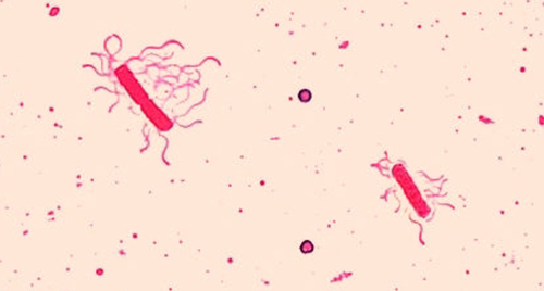
Though flagella staining is uncommon in clinical settings, the technique is commonly used by microbiologists, since the location and number of flagella can be useful in classifying and identifying bacteria in a sample. When using this technique, it is important to handle the specimen with great care; flagella are delicate structures that can easily be damaged or pulled off, compromising attempts to accurately locate and count the number of flagella.

A table of simple stains is shown. Basic stains include: methylene blue, crystal violet, malachite green, basic fuschsin, carbolfuschsin, and safranin. Basic stains stain negatively charged molecules and structures, such as nucleic acids and proteins. The outcome of this positive stain is dark cells on a light background. Acidic stains include eosine, acid fuchsin, rose Bengal, and Congo red. Acid stains stain positively charged molecules and structures such as proteins. Acidic stains can either be positive or negative stains depending on the cell’s chemistry. Negative stains include india in k and nigrosine. Negative stains stain the background, not the specimen and produce a dark background with a light specimen.
(credit “basic stains”: modification of work by Centers for Disease Control and Prevention; credit “Acidic stains”: modification of work by Roberto Danovaro, Antonio Dell’Anno, Antonio Pusceddu, Cristina Gambi, Iben Heiner, Reinhardt Mobjerg Kristensen; credit “Negative stains”: modification of work by Anh-Hue Tu)
A table of differential stains is shown. The Gram stain uses crystal violet, Gram’s iodine, ethanol (decolorizer), and safranin. The purpose of the Gram stain is to distinguish cells by cell-wall type (Gram-positive, Gram-negative). Gram-positive cells stain purple/violet. Gram-negative cells stain pink. The acid fast stain: after staining with basic fuchsin, acid-fast bacteria resist decolonization by acid-alcohol. Non-acid-fast bacteria are counterstained with methylene blue. The acid-fast stain is used to distinguish acid-fast bacteria such as M. tuberculosis, from non-acid-fast cells. Acid-fast bacteria are red; non-acid-fast cells are blue. The Endospore stain uses heat to stain endospores with malachite green (Schaeffer-Fulton procedure), then cell is washed and counterstained with safranin. The endospore stain is used to distinguish organisms with endospores from those without; used to study the endospore. Endospores appear bluish-green; other structures appear pink to red. Flagella stain: flagella are coated with a tannic acid or potassium alum mordant, then stained using either pararosaline or basic fuchsin. The flagella stain is used to view and study flagella in bacteria that have them. Flagella are visible as thin strands if present. Capsule stain: negative staining with india ink or nigrosine is used to stain the background, leaving a clear area of the cell and the capsule Counterstaining can be used to stain the cell while leaving the capsule clear. The capsule stain is used to distinguish cells with capsules from those without. Capsules appear clear or as halos if present.
(credit “Gram stain”: modification of work by Nina Parker; credit “Acid-fast stain”: modification of work by American Society for Microbiology; credit “Endospore stain”: modification of work by American Society for Microbiology; credit “Capsule stain” : modification of work by American Society for Microbiology; credit “Flagella stain”: modification of work by Centers for Disease Control and Prevention)

Preparing specimens for electron microscopy

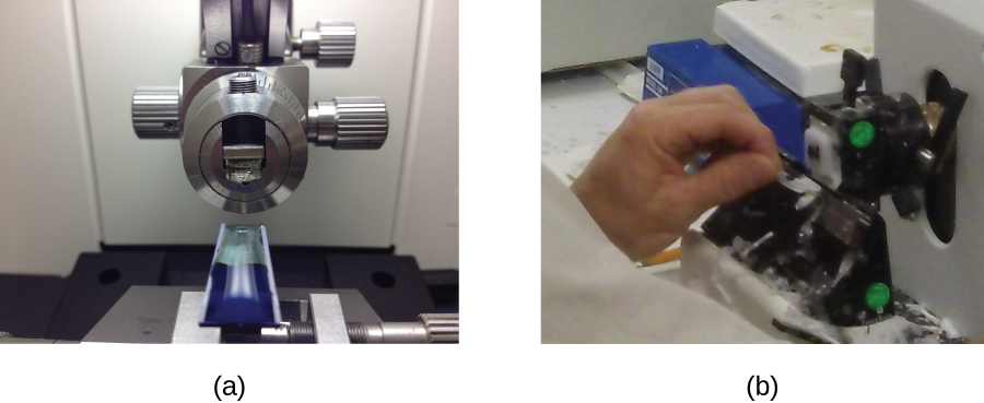
Samples to be analyzed using a TEM must have very thin sections. But cells are too soft to cut thinly, even with diamond knives. To cut cells without damage, the cells must be embedded in plastic resin and then dehydrated through a series of soaks in ethanol solutions (50%, 60%, 70%, and so on). The ethanol replaces the water in the cells, and the resin dissolves in ethanol and enters the cell, where it solidifies. Next, thin sections are cut using a specialized device called an ultramicrotome ( [link] ). Finally, samples are fixed to fine copper wire or carbon-fiber grids and stained—not with colored dyes, but with substances like uranyl acetate or osmium tetroxide, which contain electron-dense heavy metal atoms.

Photograph a shows a blue solid specimen sits below a mechanical dial. Photograph b shows a person holding a dial on a machine.
(a) An ultramicrotome used to prepare specimens for a TEM. (b) A technician uses an ultramicrotome to slice a specimen into thin sections. (credit a: modification of work by “Frost Museum”/Flickr; credit b: modification of work by U.S. Fish and Wildlife Service Northeast Region)

Questions & Answers

how does Neisseria cause meningitis
Nyibol Reply
what is microbiologist
Muhammad Reply
what is errata
Muhammad
is the branch of biology that deals with the study of microorganisms.
Ntefuni Reply
What is microbiology
Mercy Reply
studies of microbes
Louisiaste
when we takee the specimen which lumbar,spin,
Ziyad Reply
How bacteria create energy to survive?
Muhamad Reply
Bacteria doesn't produce energy they are dependent upon their substrate in case of lack of nutrients they are able to make spores which helps them to sustain in harsh environments
_Adnan
But not all bacteria make spores, l mean Eukaryotic cells have Mitochondria which acts as powerhouse for them, since bacteria don't have it, what is the substitution for it?
Muhamad
they make spores
Louisiaste
what is sporadic nd endemic, epidemic
Aminu Reply
the significance of food webs for disease transmission
Abreham
food webs brings about an infection as an individual depends on number of diseased foods or carriers dully.
Mark
explain assimilatory nitrate reduction
Esinniobiwa Reply
Assimilatory nitrate reduction is a process that occurs in some microorganisms, such as bacteria and archaea, in which nitrate (NO3-) is reduced to nitrite (NO2-), and then further reduced to ammonia (NH3).
Elkana
This process is called assimilatory nitrate reduction because the nitrogen that is produced is incorporated in the cells of microorganisms where it can be used in the synthesis of amino acids and other nitrogen products
Elkana
Examples of thermophilic organisms
Shu Reply
Give Examples of thermophilic organisms
Shu
advantages of normal Flora to the host
Micheal Reply
Prevent foreign microbes to the host
Abubakar
they provide healthier benefits to their hosts
ayesha
They are friends to host only when Host immune system is strong and become enemies when the host immune system is weakened . very bad relationship!
Mark
what is cell
faisal Reply
cell is the smallest unit of life
Fauziya
cell is the smallest unit of life
Akanni
ok
Innocent
cell is the structural and functional unit of life
Hasan
is the fundamental units of Life
Musa
what are emergency diseases
Micheal Reply
There are nothing like emergency disease but there are some common medical emergency which can occur simultaneously like Bleeding,heart attack,Breathing difficulties,severe pain heart stock.Hope you will get my point .Have a nice day ❣️
_Adnan
define infection ,prevention and control
Innocent
I think infection prevention and control is the avoidance of all things we do that gives out break of infections and promotion of health practices that promote life
Lubega
Heyy Lubega hussein where are u from?
_Adnan
en français
Adama
which site have a normal flora
ESTHER Reply
Many sites of the body have it Skin Nasal cavity Oral cavity Gastro intestinal tract
Safaa
skin
Asiina
skin,Oral,Nasal,GIt
Sadik
How can Commensal can Bacteria change into pathogen?
Sadik
How can Commensal Bacteria change into pathogen?
Sadik
all
Tesfaye
by fussion
Asiina
what are the advantages of normal Flora to the host
Micheal
what are the ways of control and prevention of nosocomial infection in the hospital
Micheal
what is inflammation
Shelly Reply
part of a tissue or an organ being wounded or bruised.
Wilfred
what term is used to name and classify microorganisms?
Micheal Reply
Binomial nomenclature
adeolu

Get Jobilize Job Search Mobile App in your pocket Now!

Get it on Google Play Download on the App Store Now


Source:  OpenStax, Microbiology. OpenStax CNX. Nov 01, 2016 Download for free at http://cnx.org/content/col12087/1.4
Google Play and the Google Play logo are trademarks of Google Inc.

Notification Switch

Would you like to follow the 'Microbiology' conversation and receive update notifications?

Ask