<< Chapter < Page Chapter >> Page >

The two suture lines seen on the top of the skull are the coronal and sagittal sutures. The coronal suture    runs from side to side across the skull, within the coronal plane of section (see [link] ). It joins the frontal bone to the right and left parietal bones. The sagittal suture    extends posteriorly from the coronal suture, running along the midline at the top of the skull in the sagittal plane of section (see [link] ). It unites the right and left parietal bones. On the posterior skull, the sagittal suture terminates by joining the lambdoid suture. The lambdoid suture    extends downward and laterally to either side away from its junction with the sagittal suture. The lambdoid suture joins the occipital bone to the right and left parietal and temporal bones.

Facial bones of the skull

The facial bones of the skull form the upper and lower jaws, the nose, nasal cavity and nasal septum, and the orbit. The facial bones include 14 bones, with six paired bones and two unpaired bones. The paired bones are the maxilla, palatine, zygomatic, nasal, lacrimal, and inferior nasal conchae bones. The unpaired bones are the vomer and mandible bones. Although classified with the brain-case bones, the ethmoid bone also contributes to the nasal septum and the walls of the nasal cavity and orbit.

Maxillary bone

The maxillary bone    , often referred to simply as the maxilla (plural = maxillae), is one of a pair that together form the upper jaw, much of the hard palate, and the base of the nose (see [link] ). Each tooth is anchored into a deep socket called an alveolus. . The hard palate    is the bony plate that forms the roof of the mouth and floor of the nasal cavity, separating the oral and nasal cavities.

Maxillary bone

This image shows the location and structure of the maxilla. A small image of the skull on the top left shows the maxilla in ochre yellow. A magnified view shows the detailed structure of the maxilla.
The maxillary bone forms the upper jaw and supports the upper teeth. Each maxilla also forms the lateral floor of each orbit and the majority of the hard palate.

Zygomatic bone

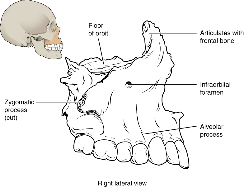
The zygomatic bone    is also known as the cheekbone. Each of the paired zygomatic bones forms much of the wall of the orbit.

Nasal bone

The nasal bone    is one of two small bones that articulate (join) with each other to form the bony base (bridge) of the nose. They also support the cartilages that form the lateral walls of the nose (see [link] ). These are the bones that are damaged when the nose is broken.

Lacrimal bone

Each lacrimal bone    is a small, rectangular bone that forms the anterior, medial wall of the orbit This bone contains the nasalacrimal canal, through which tears pass.

Vomer bone

The unpaired vomer bone, often referred to simply as the vomer, is triangular-shaped and forms the lower part of the nasal septum (see [link] ).

Mandible

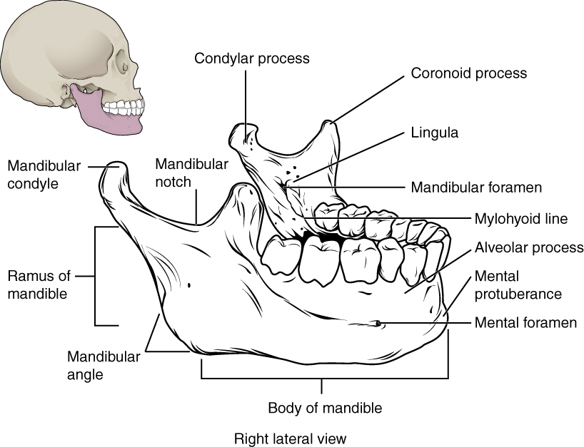
The mandible    forms the lower jaw and is the only moveable bone of the skull. At the time of birth, the mandible consists of paired right and left bones, but these fuse together during the first year to form the single U-shaped mandible of the adult skull.

Isolated mandible

This image shows the structure of the mandible. On the top left, a lateral view of the skull shows the location of the mandible in purple. A magnified image shows the right lateral view of the mandible with the major parts labeled.
The mandible is the only moveable bone of the skull.

Paranasal sinuses

The paranasal sinuses    are hollow, air-filled spaces located within certain bones of the skull ( [link] ). All of the sinuses communicate with the nasal cavity (paranasal = “next to nasal cavity”) and are lined with nasal mucosa. They serve to reduce bone mass and thus lighten the skull, and they also add resonance to the voice. This second feature is most obvious when you have a cold or sinus congestion. These produce swelling of the mucosa and excess mucus production, which can obstruct the narrow passageways between the sinuses and the nasal cavity, causing your voice to sound different to yourself and others. This blockage can also allow the sinuses to fill with fluid, with the resulting pressure producing pain and discomfort.

The paranasal sinuses are named for the skull bone that each occupies. The frontal sinus    is located just above the eyebrows, within the frontal bone (see [link] ). The largest sinus is the maxillary sinus    . These are paired and located within the right and left maxillary bones, where they occupy the area just below the orbits. The maxillary sinuses are most commonly involved during sinus infection because of their close connection to the nasal cavity. The sphenoid sinus    is a single, midline sinus. The ethmoid bone contains multiple small spaces separated by very thin bony walls. These are collectively called the ethmoid sinus .

Paranasal sinuses

This figure shows a woman’s face and the location of the paranasal sinuses. The left panel shows the anterior view of the woman’s face with the sinuses labeled. The right panel shows the lateral view of the woman’s face with the same parts labeled.
The paranasal sinuses are hollow, air-filled spaces named for the skull bone that each occupies. The most anterior is the frontal sinus, located in the frontal bone above the eyebrows. The largest are the maxillary sinuses, located in the right and left maxillary bones below the orbits. The most posterior is the sphenoid sinus, located in the body of the sphenoid bone, under the sella turcica. The ethmoid air cells are multiple small spaces located in the right and left sides of the ethmoid bone, between the medial wall of the orbit and lateral wall of the upper nasal cavity.

Hyoid bone

The hyoid bone is an independent bone that does not contact any other bone and thus is not part of the skull ( [link] ). It is a small U-shaped bone located in the upper neck near the mandible, with the tips of the “U” pointing backwards. The hyoid serves as the base for the tongue above, and is attached to the larynx and the pharynx .

Hyoid bone

In this image, the location and structure of the hyoid bone are shown. The top panel shows a person’s face and neck, with the hyoid bone highlighted in grey. The middle panel shows the anterior view and the bottom panel shows the right anterior view.
The hyoid bone is located in the upper neck and does not join with any other bone. It provides attachments for muscles that act on the tongue, larynx, and pharynx.

References

Centers for Disease Control and Prevention (US). Injury prevention and control: traumatic brain injury [Internet]. Atlanta, GA; [cited 2013 Mar 18]. Available from: (External Link) .

Questions & Answers

I'm interested in biological psychology and cognitive psychology
Tanya Reply
what does preconceived mean
sammie Reply
physiological Psychology
Nwosu Reply
How can I develope my cognitive domain
Amanyire Reply
why is communication effective
Dakolo Reply
Communication is effective because it allows individuals to share ideas, thoughts, and information with others.
effective communication can lead to improved outcomes in various settings, including personal relationships, business environments, and educational settings. By communicating effectively, individuals can negotiate effectively, solve problems collaboratively, and work towards common goals.
it starts up serve and return practice/assessments.it helps find voice talking therapy also assessments through relaxed conversation.
miss
Every time someone flushes a toilet in the apartment building, the person begins to jumb back automatically after hearing the flush, before the water temperature changes. Identify the types of learning, if it is classical conditioning identify the NS, UCS, CS and CR. If it is operant conditioning, identify the type of consequence positive reinforcement, negative reinforcement or punishment
Wekolamo Reply
please i need answer
Wekolamo
because it helps many people around the world to understand how to interact with other people and understand them well, for example at work (job).
Manix Reply
Agreed 👍 There are many parts of our brains and behaviors, we really need to get to know. Blessings for everyone and happy Sunday!
ARC
A child is a member of community not society elucidate ?
JESSY Reply
Isn't practices worldwide, be it psychology, be it science. isn't much just a false belief of control over something the mind cannot truly comprehend?
Simon Reply
compare and contrast skinner's perspective on personality development on freud
namakula Reply
Skinner skipped the whole unconscious phenomenon and rather emphasized on classical conditioning
war
explain how nature and nurture affect the development and later the productivity of an individual.
Amesalu Reply
nature is an hereditary factor while nurture is an environmental factor which constitute an individual personality. so if an individual's parent has a deviant behavior and was also brought up in an deviant environment, observation of the behavior and the inborn trait we make the individual deviant.
Samuel
I am taking this course because I am hoping that I could somehow learn more about my chosen field of interest and due to the fact that being a PsyD really ignites my passion as an individual the more I hope to learn about developing and literally explore the complexity of my critical thinking skills
Zyryn Reply
good👍
Jonathan
and having a good philosophy of the world is like a sandwich and a peanut butter 👍
Jonathan
generally amnesi how long yrs memory loss
Kelu Reply
interpersonal relationships
Abdulfatai Reply
What would be the best educational aid(s) for gifted kids/savants?
Heidi Reply
treat them normal, if they want help then give them. that will make everyone happy
Saurabh
Got questions? Join the online conversation and get instant answers!
Jobilize.com Reply

Get Jobilize Job Search Mobile App in your pocket Now!

Get it on Google Play Download on the App Store Now


Source:  OpenStax, Skeletal system. OpenStax CNX. Apr 17, 2015 Download for free at https://legacy.cnx.org/content/col11779/1.1
Google Play and the Google Play logo are trademarks of Google Inc.

Notification Switch

Would you like to follow the 'Skeletal system' conversation and receive update notifications?

Ask