<< Chapter < Page Chapter >> Page >

Body symmetry

Animals may be asymmetrical, radial, or bilateral in form ( [link] ). Asymmetrical animals are animals with no pattern or symmetry; an example of an asymmetrical animal is a sponge ( [link] a ). An organism with radial symmetry    ( [link] b ) has a longitudinal (up-and-down) orientation: Any plane cut along this up–down axis produces roughly mirror-image halves. An example of an organism with radial symmetry is a sea anemone.

Illustration a shows an asymmetrical sponge with a tube-like body and a growth off to one side. Illustration b shows a sea anemone with a tube-like, radially symmetrical body. Tentacles grow from the top of the tube. Three vertical planes arranged 120 degrees apart dissect the body. The half of the body on one side of each plane is a mirror image of the body on the other side. Illustration c shows a goat with a bilaterally symmetrical body. A plane runs from front to back through the middle of the goat, dissecting the body into left and right halves, which are mirror images of each other. The top part of the goat is defined as dorsal, and the bottom part is defined as ventral. The front of the goat is defined as anterior, and the back is defined as posterior.
Animals exhibit different types of body symmetry. The (a) sponge is asymmetrical and has no planes of symmetry, the (b) sea anemone has radial symmetry with multiple planes of symmetry, and the (c) goat has bilateral symmetry with one plane of symmetry.

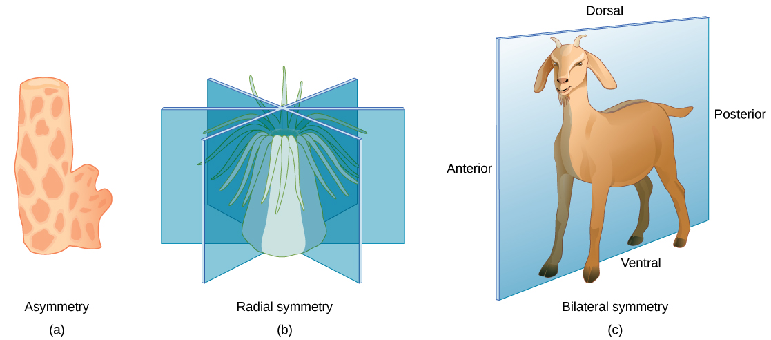
Bilateral symmetry is illustrated in [link] c using a goat. The goat also has upper and lower sides to it, but they are not symmetrical. A vertical plane cut from front to back separates the animal into roughly mirror-image right and left sides. Animals with bilateral symmetry also have a “head” and “tail” (anterior versus posterior) and a back and underside (dorsal versus ventral).

Concept in action

Watch this video to see a quick sketch of the different types of body symmetry.

Layers of tissues

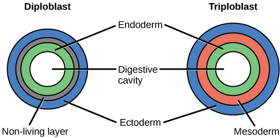
Most animal species undergo a layering of early tissues during embryonic development. These layers are called germ layers . Each layer develops into a specific set of tissues and organs. Animals develop either two or three embryonic germs layers ( [link] ). The animals that display radial symmetry develop two germ layers, an inner layer (endoderm) and an outer layer (ectoderm). These animals are called diploblasts . Animals with bilateral symmetry develop three germ layers: an inner layer (endoderm), an outer layer (ectoderm), and a middle layer (mesoderm). Animals with three germ layers are called triploblasts .

The left illustration shows the two embryonic germ layers of a diploblast. The inner layer is the endoderm, and the outer layer is the ectoderm. Sandwiched between the endoderm and the ectoderm is a non-living layer. The right illustration shows the three embryonic germ layers of a triploblast. Like the diploblast, the triploblast has an inner endoderm and an outer ectoderm. Sandwiched between these two layers is a living mesoderm.
During embryogenesis, diploblasts develop two embryonic germ layers: an ectoderm and an endoderm. Triploblasts develop a third layer—the mesoderm—between the endoderm and ectoderm.

Presence or absence of a coelom

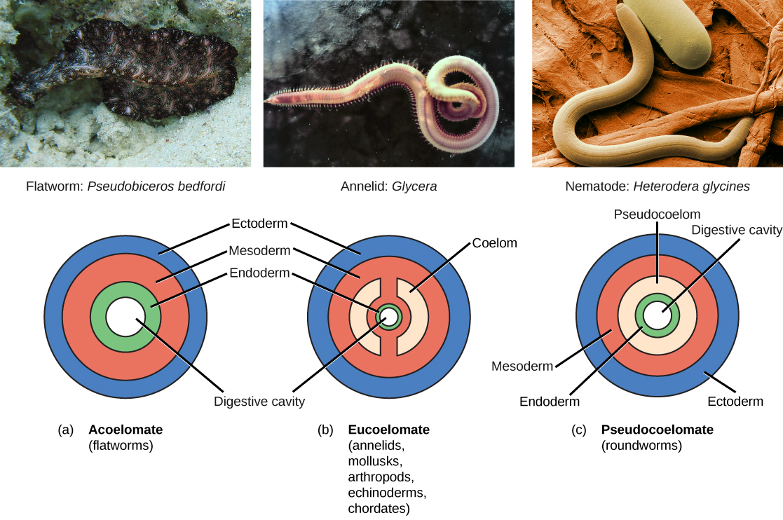
Triploblasts may develop an internal body cavity derived from mesoderm, called a coelom    (pr. see-LŌM). This epithelial-lined cavity is a space, usually filled with fluid, which lies between the digestive system and the body wall. It houses organs such as the kidneys and spleen, and contains the circulatory system. Triploblasts that do not develop a coelom are called acoelomates , and their mesoderm region is completely filled with tissue, although they have a gut cavity. Examples of acoelomates include the flatworms. Animals with a true coelom are called eucoelomates (or coelomates) ( [link] ). A true coelom arises entirely within the mesoderm germ layer. Animals such as earthworms, snails, insects, starfish, and vertebrates are all eucoelomates. A third group of triploblasts has a body cavity that is derived partly from mesoderm and partly from endoderm tissue. These animals are called pseudocoelomates . Roundworms are examples of pseudocoelomates. New data on the relationships of pseudocoelomates suggest that these phyla are not closely related and so the evolution of the pseudocoelom must have occurred more than once ( [link] ). True coelomates can be further characterized based on features of their early embryological development.

Part a shows the body plan of acoelomates, including flatworms. Acoelomates have a central digestive cavity. Outside this digestive cavity are three tissue layers: an inner endoderm, a central mesoderm, and an outer ectoderm. The photo shows a swimming flatworm, which has the appearance of a frilly black and pink ribbon. Part b shows the body plan of eucoelomates, which include annelids, mollusks, arthropods, echinoderms, and chordates. Eucoelomates have the same tissue layers as acoelomates, but a cavity called a coelom exists within the mesoderm. The coelom is divided into two symmetrical parts that are separated by two spokes of mesoderm. The photo shows a swimming annelid known as a bloodworm. The bloodworm has a tubular body that is tapered at each end. Numerous appendages radiate from either side. Part c shows the body plan of pseudocoelomates, which include roundworms. Like the acoelomates and eucoelomates, the pseudocoelomates have an endoderm, a mesoderm, and an ectoderm. However, in pseudocoelomates, a pseudocoelom separates the endoderm from the mesoderm. The photo shows a roundworm, or nematode, which has a tubular body.
Triploblasts may be acoelomates, eucoelomates, or pseudocoelomates. Eucoelomates have a body cavity within the mesoderm, called a coelom, which is lined with mesoderm tissue. Pseudocoelomates have a similar body cavity, but it is lined with mesoderm and endoderm tissue. (credit a: modification of work by Jan Derk; credit b: modification of work by NOAA; credit c: modification of work by USDA, ARS)

Protostomes and deuterostomes

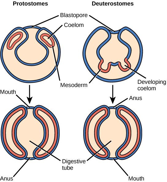
Bilaterally symmetrical, triploblastic eucoelomates can be divided into two groups based on differences in their early embryonic development. Protostomes include phyla such as arthropods, mollusks, and annelids. Deuterostomes include the chordates and echinoderms. These two groups are named from which opening of the digestive cavity develops first: mouth or anus. The word protostome comes from Greek words meaning “mouth first,” and deuterostome originates from words meaning “mouth second” (in this case, the anus develops first). This difference reflects the fate of a structure called the blastopore ( [link] ), which becomes the mouth in protostomes and the anus in deuterostomes. Other developmental characteristics differ between protostomes and deuterostomes, including the mode of formation of the coelom and the early cell division of the embryo.

The illustration compares the development of protostomes and deuterostomes. In both protostomes and deuterostomes, the gastrula, which resembles a hollow ball of cells, contains an indentation called a blastopore. In protostomes, two circular layers of mesoderm form inside the gastrula, containing the coelom. As the protostome develops, the mesoderm grows and fuses with the gastrula cell layer. The blastopore becomes the mouth, and a second opening forms opposite the mouth, which becomes the anus. In deuterostomes, two groups of gastrula cells in the blastopore grow inward to form the mesoderm. As the deuterostome develops, the mesoderm pinches off and fuses, forming a second body cavity. The body plan of the deuterostome at this stage looks very similar to that of the protostome, but the blastopore becomes the anus, and the second opening becomes the mouth.
Eucoelomates can be divided into two groups, protostomes and deuterostomes, based on their early embryonic development. Two of these differences include the origin of the mouth opening and the way in which the coelom is formed.

Section summary

Animals constitute a diverse kingdom of organisms. Although animals range in complexity from simple sea sponges to human beings, most members share certain features. Animals are eukaryotic, multicellular, heterotrophic organisms that ingest their food and usually develop into motile creatures with a fixed body plan. Most members of the animal kingdom have differentiated tissues of four main classes—nervous, muscular, connective, and epithelial—that are specialized to perform different functions. Most animals reproduce sexually, leading to a developmental sequence that is relatively similar across the animal kingdom.

Organisms in the animal kingdom are classified based on their body morphology and development. True animals are divided into those with radial versus bilateral symmetry. Animals with three germ layers, called triploblasts, are further characterized by the presence or absence of an internal body cavity called a coelom. Animals with a body cavity may be either coelomates or pseudocoelomates, depending on which tissue gives rise to the coelom. Coelomates are further divided into two groups called protostomes and deuterostomes, based on a number of developmental characteristics.

Art connection

[link] Which of the following statements is false?

  1. Eumetazoa have specialized tissues and Parazoa do not.
  2. Both acoelomates and pseudocoelomates have a body cavity.
  3. Chordates are more closely related to echinoderms than to rotifers according to the figure.
  4. Some animals have radial symmetry, and some animals have bilateral symmetry.

[link] B

Got questions? Get instant answers now!

Questions & Answers

what Is taxonom
Don Reply
Taxonom is the the laws and principles guiding the classification of living things either plant 🌿 or animal
Samuel
protein synthesis began with what?
Kesselly Reply
What structures does a plant cell have that an animal cell does not hav
Martin Reply
A plant cell has a cell wall, a large central vacuole, and chloroplasts, which an animal cell does not have.
AI-Robot
what is the function of the cytoskeleton?
Martin
truly. Al-robot
Mark
why is it advantageous for the cell membrane to be fluid in nature?
Martin Reply
The fluid nature of the cell membrane allows for flexibility and dynamic changes in its structure. This allows for the movement of molecules and ions in and out of the cell, as well as the ability to respond to external stimuli. The fluidity of the membrane also allows for the formation of specialized structures such as receptor proteins, which are important for cell-cell communication.
AI-Robot
What is used as a visual marker for successful gene transfer into transgenic animals?
rhonda Reply
Parts of the body involved in homeostasis
Favour Reply
liver
Kirk
A collection of similar-typed cells working together to perform a specific function is called what?
Rachel Reply
tissue
Van
which structure consists of a phospholipid bilayers and it is known as the mosaic model?
Jamar Reply
cell membrane
Abanoub
what is diffusion
Patricia Reply
a passive process of transport
Jamar
Random movement of gas molecule from region of high concentration to a low concentration.
praise
10 difference between male and female reproductive system
Ina Reply
if a phase is omitted ,what will happen to the cell
Ewemoje Reply
discribe advantages and disadvantages of asexual and sexual reproduction
basi Reply
Compared to separate sexes and assuming self-fertilizing is not possible, what might be one advantage and one disadvantage to hermaphroditism?
Gift Reply
is nature of their environment among
basi
cool 😎
Precious
why are the laws of thermodynamics considered laws of nature and not scientific theories
Yolonda Reply
archea were given their own separate domain because they are?
Yolonda
which of the following is a basic component of all of the others?
Yolonda
which of the following organization levels is the least inclusive
Yolonda
which cell feature is absent in bacterial cells
Yolonda
which metric movies the base unit of measurement by one thousandth (0.001)?
Yolonda
What is biology?
Blessing Reply
List the branches of biology
Blessing
List the branches of biology
Blessing
List the branches of biology
Blessing
List the branches of biology
Blessing
Ecology,Anatomy,genetics,biophysics,biochemistry,microbiology and biotechnology.
praise

Get Jobilize Job Search Mobile App in your pocket Now!

Get it on Google Play Download on the App Store Now


Source:  OpenStax, Concepts of biology. OpenStax CNX. Feb 29, 2016 Download for free at http://cnx.org/content/col11487/1.9
Google Play and the Google Play logo are trademarks of Google Inc.

Notification Switch

Would you like to follow the 'Concepts of biology' conversation and receive update notifications?

Ask