<< Chapter < Page Chapter >> Page >

Art connection

The kidney is shaped like a kidney bean standing on end. Two layers, the outer renal fascia and an inner capsule, cover the outside of the kidney. The inside of the kidney consists of three layers: the outer cortex, the middle medulla and the inner renal pelvis. The renal pelvis is flush with the concave side of the kidney, and empties into the ureter, a tube that runs down outside the concave side of the kidney. Nine renal pyramids are embedded in the medulla, which is the thickest kidney layer. Each renal pyramid is teardrop-shaped, with the narrow end facing the renal pelvis. The renal artery and renal vein enter the concave part of the kidney, just above the ureter. The renal artery and renal vein branch into arterioles and venuoles, respectively, which extend into the kidney and branch into capillaries in the cortex.
The internal structure of the kidney is shown. (credit: modification of work by NCI)

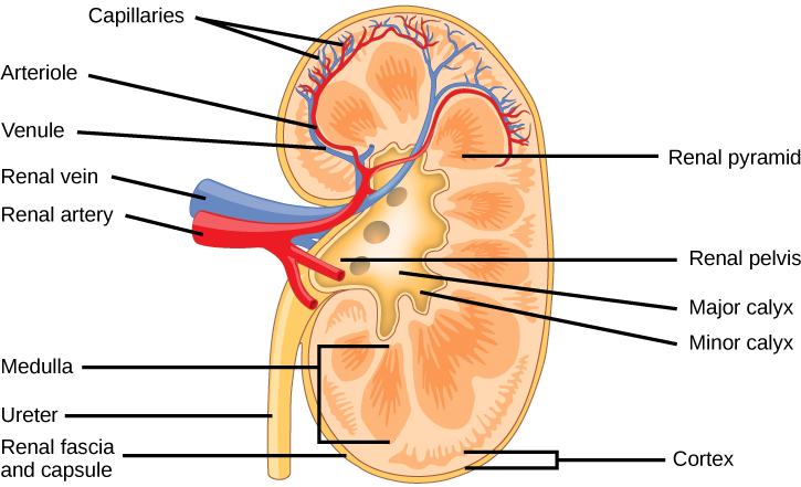
Because the kidney filters blood, its network of blood vessels is an important component of its structure and function. The arteries, veins, and nerves that supply the kidney enter and exit at the renal hilum. Renal blood supply starts with the branching of the aorta into the renal arteries and ends with the exiting of the renal veins to join the inferior vena cava    , which transports blood back to the right atrium of the heart. The renal arteries split multiple times to form other blood vessels before branching into numerous afferent arterioles, and then enter the capillaries supplying the nephrons.

As mentioned previously, the functional unit of the kidney is the nephron, illustrated in [link] . Each kidney is made up of over one million nephrons that dot the renal cortex. A nephron consists of three parts—a renal corpuscle    , a renal tubule    , and the associated capillary network.

Art connection

Illustration shows the nephron, a tube-like structure that begins in the kidney cortex. Here, arterioles converge in a bulb-like structure called the glomerulus, which is partly surrounded by a Bowman’s capsule. Afferent arterioles enter the glomerulus, and efferent arterioles leave. The glomerulus empties into the proximal convoluted tubule. A long loop, called the loop of Henle, extends from the proximal convoluted tubule to the inner medulla of the kidney, and then back out to the cortex. There, the loop of Henle joins a distal convoluted tubule. The distal convoluted tubule joins a collecting duct, which travels from the medulla back into the cortex, toward the center of the kidney. Eventually, the contents of the renal pyramid empty into the renal pelvis, and then the ureter.
The nephron is the functional unit of the kidney. The glomerulus and convoluted tubules are located in the kidney cortex, while collecting ducts are located in the pyramids of the medulla. (credit: modification of work by NIDDK)

Renal corpuscle

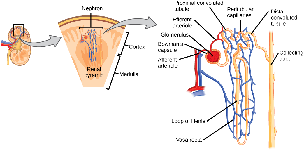
The renal corpuscle, located in the renal cortex, is made up of a network of capillaries known as the glomerulus and the capsule, a cup-shaped chamber that surrounds it, called the glomerular or Bowman's capsule    .

Renal tubule

The renal tubule is a long and convoluted structure that emerges from the glomerulus and can be divided into three parts based on function. The first part is called the proximal convoluted tubule (PCT)    due to its proximity to the glomerulus. The second part is called the loop of Henle    , because it forms a loop (with descending and ascending limbs ). The third part of the renal tubule is called the distal convoluted tubule (DCT)    . The DCT, which is the last part of the nephron, connects and empties its contents into collecting ducts. The urine will ultimately move into the renal pelvis and then into the ureters.

Capillary network within the nephron

The capillary network that originates from the renal arteries supplies the nephron with blood that needs to be filtered. The branch that enters the glomerulus is called the afferent arteriole    . The branch that exits the glomerulus is called the efferent arteriole    . Within the glomerulus, the network of capillaries is called the glomerular capillary bed. Once the efferent arteriole exits the glomerulus, it forms the peritubular capillary network    , which surrounds and interacts with parts of the renal tubule.

Go to this website to see another section of the kidney and to explore an animation of the workings of nephrons.

Kidney function and physiology

Kidneys filter blood in a three-step process. First, the nephrons filter blood that runs through the capillary network in the glomerulus. Almost all solutes, except for proteins, are filtered out into the glomerulus by a process called glomerular filtration    . Second, the filtrate is collected in the renal tubules. Most of the solutes get reabsorbed in the PCT by a process called tubular reabsorption    . In the loop of Henle, the filtrate continues to exchange solutes and water with the peritubular capillary network. Water is also reabsorbed during this step. Then, additional solutes and wastes are secreted into the kidney tubules during tubular secretion    , which is, in essence, the opposite process to tubular reabsorption. The collecting ducts collect filtrate coming from the nephrons and this filtrate, called urine, will be transported into the renal pelvis and then to the ureters. This entire process is illustrated in [link] .

Questions & Answers

Explain the following terms . (1) Abiotic factors in an ecosystem
Nomai Reply
Abiotic factors are non living components of ecosystem.These include physical and chemical elements like temperature,light,water,soil,air quality and oxygen etc
Qasim
what is biology
daniel Reply
what is diffusion
Emmanuel Reply
passive process of transport of low-molecular weight material according to its concentration gradient
AI-Robot
what is production?
Catherine
Pathogens and diseases
how did the oxygen help a human being
Achol Reply
how did the nutrition help the plants
Achol Reply
Biology is a branch of Natural science which deals/About living Organism.
Ahmedin Reply
what is phylogeny
Odigie Reply
evolutionary history and relationship of an organism or group of organisms
AI-Robot
ok
Deng
what is biology
Hajah Reply
cell is the smallest unit of the humanity biologically
Abraham
what is biology
Victoria Reply
what is biology
Abraham
HOW CAN MAN ORGAN FUNCTION
Alfred Reply
the diagram of the digestive system
Assiatu Reply
allimentary cannel
Ogenrwot
How does twins formed
William Reply
They formed in two ways first when one sperm and one egg are splited by mitosis or two sperm and two eggs join together
Oluwatobi
what is respiration
Deborah
what is genetics
Josephine Reply
Genetics is the study of heredity
Misack
how does twins formed?
Misack
What is manual
Hassan Reply
Got questions? Join the online conversation and get instant answers!
Jobilize.com Reply

Get Jobilize Job Search Mobile App in your pocket Now!

Get it on Google Play Download on the App Store Now


Source:  OpenStax, Human biology. OpenStax CNX. Dec 01, 2015 Download for free at http://legacy.cnx.org/content/col11903/1.3
Google Play and the Google Play logo are trademarks of Google Inc.

Notification Switch

Would you like to follow the 'Human biology' conversation and receive update notifications?

Ask