Viruses are classified in several ways: by factors such as their core content (
[link] and
[link] ), the structure of their capsids, and whether they have an outer envelope. The type of genetic material (DNA or RNA) and its structure (single- or double-stranded, linear or circular, and segmented or non-segmented) are used to classify the virus core structures.
Non-segmented: genome consists of a single segment of genetic material
Segmented: genome is divided into multiple segments
Parainfluenza viruses
Influenza viruses
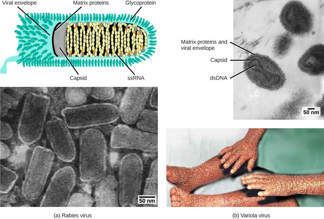
Viruses are classified based on their core genetic material and capsid design. (a) Rabies virus has a single-stranded RNA (ssRNA) core and an enveloped helical capsid, whereas (b) variola virus, the causative agent of smallpox, has a double-stranded DNA (dsDNA) core and a complex capsid. Rabies transmission occurs when saliva from an infected mammal enters a wound. The virus travels through neurons in the peripheral nervous system to the central nervous system where it impairs brain function, and then travels to other tissues. The virus can infect any mammal, and most die within weeks of infection. Smallpox is a human virus transmitted by inhalation of the variola virus, localized in the skin, mouth, and throat, which causes a characteristic rash. Before its eradication in 1979, infection resulted in a 30–35 percent mortality rate. (credit “rabies diagram”: modification of work by CDC; “rabies micrograph”: modification of work by Dr. Fred Murphy, CDC; credit “small pox micrograph”: modification of work by Dr. Fred Murphy, Sylvia Whitfield, CDC; credit “smallpox photo”: modification of work by CDC; scale-bar data from Matt Russell)
Viruses can also be classified by the design of their capsids (
[link] and
[link] ). Capsids are classified as naked icosahedral, enveloped icosahedral, enveloped helical, naked helical, and complex (
[link] and
[link] ). The type of genetic material (DNA or RNA) and its structure (single- or double-stranded, linear or circular, and segmented or non-segmented) are used to classify the virus core structures (
[link] ).
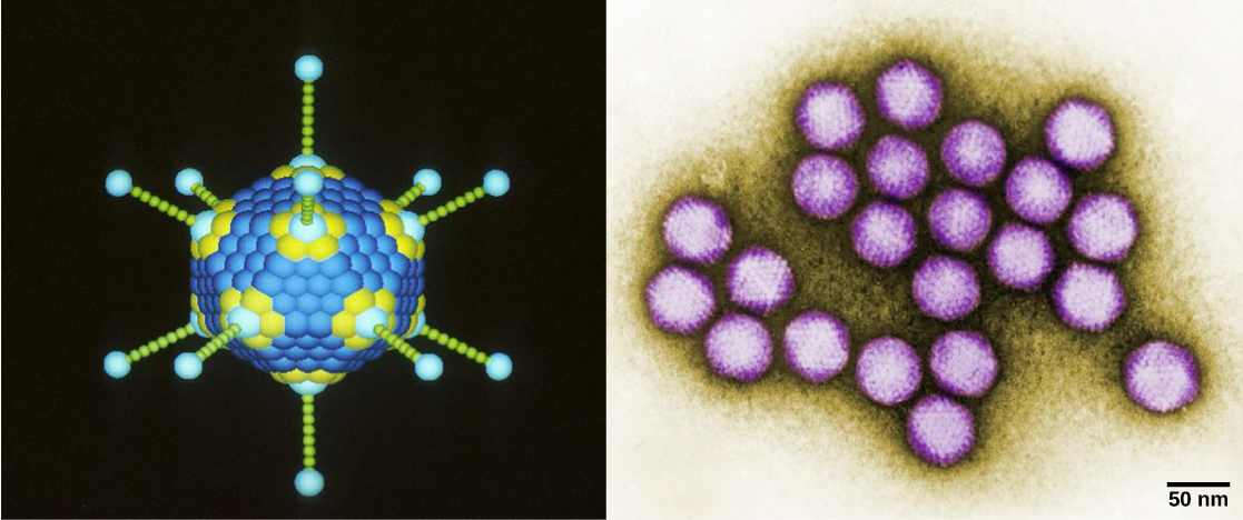
Adenovirus (left) is depicted with a double-stranded DNA genome enclosed in an icosahedral capsid that is 90–100 nm across. The virus, shown clustered in the micrograph (right), is transmitted orally and causes a variety of illnesses in vertebrates, including human eye and respiratory infections. (credit “adenovirus”: modification of work by Dr. Richard Feldmann, National Cancer Institute; credit “micrograph”: modification of work by Dr. G. William Gary, Jr., CDC; scale-bar data from Matt Russell)
the study of living organisms and their interactions with one another and their environment.
Wine
discuss the biological phenomenon and provide pieces of evidence to show that it was responsible for the formation of eukaryotic organelles in an essay form
advantage of electronic microscope is easily and clearly while disadvantage is dangerous because its electronic. advantage of light microscope is savely and naturally by sun while disadvantage is not easily,means its not sharp and not clear
Abdullahi
cell theory state that every organisms composed of one or more cell,cell is the basic unit of life
Abdullahi
is like gone fail us
DENG
cells is the basic structure and functions of all living things
A scanning electron microscope (SEM) is ideal for situations requiring high-resolution imaging of surfaces. It is commonly used in materials science, biology, and geology to examine the topography and composition of samples at a nanoscale level. SEM is particularly useful for studying fine details,